
3 2D, 3D, and 4D Ultrasound Imaging
3.1 Learning Objectives
After reviewing this chapter, you should be able to do the following:
- Define and explain the mode of operation of two-dimensional (2D), three-dimensional (3D), and four-dimensional (4D) ultrasound imaging.
- Compare and contrast the advantages and disadvantages of 2D, 3D, and 4D ultrasound imaging in medical applications.
- Evaluate the diagnostic benefits of 3D/4D ultrasound imaging over 2D imaging in different medical fields, such as obstetrics, gynecology, and cardiology.
- Analyze the limitations of 3D/4D ultrasound imaging in medical applications, including technical, clinical, and ethical considerations.
- Describe the recent improvements in technology, image quality, and clinical ease of use that have contributed to the expanding applications of 3D/4D ultrasound imaging in patient care.
- Critically assess the current use and potential future directions of 3D/4D ultrasound imaging in clinical practice, research, and education.
- Discuss the ethical implications of 3D/4D ultrasound imaging, such as fetal imaging for nonmedical purposes, and the impact of imaging on patient autonomy and decision-making.
3.2 Introduction
Medical ultrasound continues to make significant contributions to patient care by providing anatomical information needed by clinicians to make critical decisions. While 3D ultrasound has been commonly used in cardiology, obstetrics, and gynecology, its applications continue to expand as essential technological improvements, image quality, and clinical ease of use increase. In this section, we discuss the modes of operation of 2D, 3D, and 4D ultrasound imaging and their current diagnostic benefits and limitations in the medical field. First, we will consider the advantages of 3D/4D over 2D imaging.
3.3 Using 2D Versus 3D and 4D Ultrasound
Two-dimensional ultrasound is considered a standard or conventional imaging technique. In 2D scanning, a series of thin slices make up an image, and only one slice can be seen at a time.
Three- and four-dimensional clinical ultrasounds have been around for nearly 25 years. However, their use has lagged behind that of computerized tomography (CT) and magnetic resonance imaging (MRI) due to the difficulty in rendering the data in 3D.[1] However, ultrasound equipment’s increasing computing power has helped resolve complex signal processing tasks needed to render 3D ultrasound data.
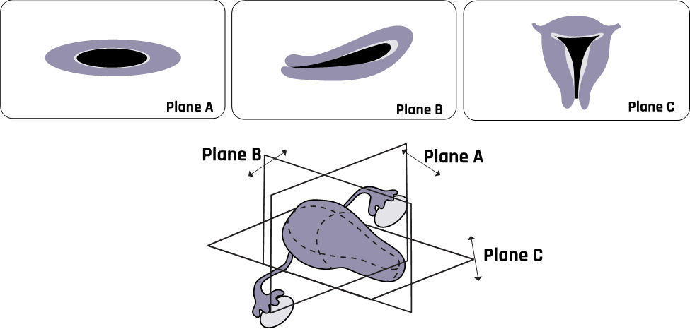
Three-dimensional ultrasound is based on the same principles of operation as 2D ultrasound but has an added position-sensing component to produce the effect of a 3D image, as illustrated in Figure 3-1. In 3D ultrasound imaging, echoes are used to form real-like realistic volume images.
A 4D ultrasound shows 3D ultrasound images in motion. Two-dimensional ultrasound images are commonly used because they are less expensive than 3D or 4D. However, many centers now use 3D/4D ultrasound. The most common area of use has been fetal cardiovascular scanning. The 3D/4D technology offers real-time motion-gated cardiac scanning.
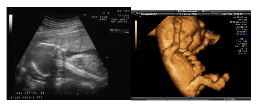
A 3D/4D ultrasound offers expecting parents a memorable lifetime opportunity to see the features of their unborn babies. Figure 3-2 shows 2D grayscale and 3D colored images.
3.4 Accuracy and Repeatability
Acquiring qualitative and quantitative sonographic volume data such as multiplanar imaging, surface and volume rendering, and semiautomated volume calculation gives 3D/4D an added advantage over 2D imaging. Virtual planes provide extra information that cannot be viewed with a standard 2D technique.[2] The American Institute of Ultrasound in Medicine (AIUM) notes that the ability to take images in any plane in real time has enormous potential use in medical diagnosis.[3] Another advantage is that 3D ultrasound can provide measurements in three planes with acceptable reliability.
3.5 Equipment Design and Image Acquisition
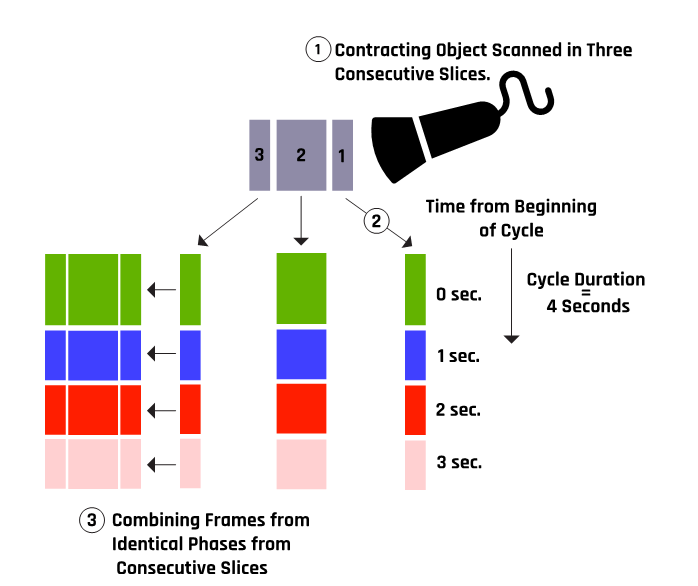
A 3D ultrasound uses a series of 2D images covering a volume of a particular area. This allows the images to be rotated and displayed in different orientations. When displayed in real time, or live, they form a 4D ultrasound. Volume acquisition is achieved by using an array of transducers consisting of many 2D frames, one behind the other. Automated mathematical algorithms are then used to process the volume data to produce the desired image, as illustrated in Figure 3-3.
The reconstruction in Figure 3-3 occurs in a matter of seconds such that the spatio-temporal imaging correlation (STIC) acquisition is completed in the presence of the patient. The STIC acquisition mode can be combined with B-mode, color, and power Doppler. From a good STIC acquisition, a sequential plane may be viewed in corresponding transverse and longitudinal planes at any time point, simultaneously.
The B-flow image is a live grayscale depiction of blood flow and cardiac chambers. When applied to 3D fetal echocardiography, the B-flow image shows blood flow in the heart and great vessels in real time.[4]
3.6 Display and Analysis
Four-dimensional imaging involves a 3D image moving in real time, created from a volume of data that allows the live reconstruction of images in different planes and renderings. Rendering is taking voxel-based data and converting it into a viewable image with added depth. Three- and four-dimensional imaging function by reconstructing an image from multiple 2D planes.
3.7 Areas of Applications
The 3D color and power Doppler ultrasound technology can be used in vascular abnormality assessments such as the fetal placental cord and pulmonary vessels. Three-dimensional ultrasound is not yet widely used in many routine medical procedures but has been widely used in gynecology, obstetrics, and biopsy.
In gynecology, 3D ultrasound can be used to
- examine and assess the uterus,
- screen for ectopic pregnancies,
- locate intrauterine devices,
- examine the ovaries, and
- perform interventional procedures for patients with infertility.[5]
In obstetrics, fetal 3D ultrasound can be used to
- examine the nasal bone, ears, and central nervous system;
- evaluate rib and lung volumes;
- screen for spinal cord, placenta, and vertebral abnormalities;
- map abnormalities in multiple gestations; and
- perform fetal cardiac scanning.
The application of 3D/4D ultrasound in biopsy allows for needle tracking in multiple planes simultaneously and imaging of the morphology and proximity of local anesthetic spread around the target nerves.[6] Other specialties in which 3D/4D ultrasound can be applied include dermatology and ophthalmology.
3.8 Advantages of 3D/4D Ultrasound Over 2D Ultrasound
In 2D ultrasound imaging, a series of noncontinuous and presumably representative sections of the imaged organ is used to visualize the anatomy of the organ. Repeated examination is required in order to reconstruct cross sections visually. The absence of quantitative spatial documentation means clinicians must rely on image labels and trust the acquisition technique. Repeated examinations may not produce exact image planes, making comparing serial exams difficult.[7] In addition, every time an organ is examined, it’s likely to give rise to varying measurements of a specific organ or structure, as the imaging process is highly operator-dependent. Volumetric (3D) ultrasound allows volumetric acquisition of anatomic data, which may be needed to examine internal organs such as the liver, gallbladder, gallstones, or kidneys.
In 3D/4D, volume data are obtained in a single image, allowing the operator to view any plane in the volume. The multiplanar rendering mode options also allow for viewing images in real-time motion. Three-dimensional imaging is helpful in examining contours such as those in the facial area, the heart chambers, and the valves. The 4D ultrasound provides a motion video of the 3D structure in real time. Together, 3D and 4D ultrasound have been quite successful in examining the inside or outside of organs, nodules, cysts, or tumors. The 3D ultrasound also offers a more comprehensive image of anatomical structures and pathological conditions. A 3D echocardiography can provide estimates of ventricular volume and function. Comparing 2D and 4D, qualitative 4D ultrasound is considered superior to 2D in real time. However, 4D is inferior to 2D ultrasound for quantitative analysis of movements. For example, 4D evaluation of complex fetal facial activity and expression is better than that of 2D.[8]
Hence 3D and 4D ultrasound imaging allow more accessible and more rapid screening than 2D ultrasound imaging. Irregularly shaped organ volumes can be more accurately visualized using 3D than 2D imaging ultrasound. In addition, 3D measurements are more reproducible than 2D ultrasound and provide the option to rotate the volume, which is often necessary to allow optimal visualization of the geometric structure of the organ under examination and determine the position of the structure in the volume.[9]
3.9 Handheld Ultrasound Devices
The market for small, portable ultrasound machines has flourished over the past decade. They have gained popularity and significant usefulness in the medical sector, especially in the emergency medicine department. There are two specific reasons for this popularity: (1) they are smaller/lighter and portable, which makes the whole process of scanning the patients quicker, more accessible, and safer at the same time, and (2) their portability, ease of use, connectivity, and cleanability make them ideal tools for diverse care settings. Conventional large-sized ultrasound machines are demanding and come with certain limits. For instance, these larger machines are laborious and challenging to sterilize thoroughly in comparison to small handheld machines. Also, a physician performing any intervention with this machine usually requires an extra pair of hands to help set it up and obtain clinical images.
Both laptop and handheld models of portable ultrasound systems are available. There are also a few models that are intermediate between these two. Therefore, portable ultrasound machines can be broadly divided into three categories. The first category is the large, laptop-sized device weighing 12 to 15 pounds. An example of such a device is GE HealthCare’s LOGIQTM e portable ultrasound. The second category involves a smaller device, almost the size of a smaller laptop and weighing about 6 to 10 pounds. An example of such a device is the Sonosite Edge machine.
With technological innovations, even smaller and lighter ultrasound devices with better image quality are now available (such as those manufactured by GE HealthCare, Siemens, and Philips). The third category is the handheld ultrasound device that weighs under a pound. Philips’s Lumify is an example of one such device; it costs around $6,000 to buy or $2,300 per year to lease. This device operates through a simple USB connection of the transducer to a compatible device such as a smartphone. GE HealthCare’s Vscan is a pocket-size ultrasound device that provides real-time gray anatomic and color flow images. It is optimized for physicians to quickly inspect the heart, abdominal organs, and urinary bladder. It can provide insights into areas of obstetrics and gynecology, pleural fluid and motion detection, and pediatrics. A recently extended version of the Vscan handheld ultrasound device can be used to help confirm and monitor the progression of acute respiratory diseases like COVID-19.
3.10 Limitations and Challenges
While 3D/4D ultrasounds show success in some cases, they have some limitations in their clinical applications. Three-dimensional renderings have demonstrated impressive results in some areas; the interfaces are complex and difficult to interpret. Other significant issues raised by the AIUM[10] are that the protocols and manipulation techniques are not standardized across manufacturers with respect to the terminology of functions and the display, therefore requiring time and effort to perfect operator skill and accuracy. Standardization is also needed in image orientation. Another challenge is that 3D/4D does not solve poor 2D image problems for several reasons: (1) artifacts and orientation can be confusing, and (2) resolution decreases in reconstructed images such that in some cases, 3D/4D images may be inferior to 2D images. Currently, 2D fetal echocardiography with color Doppler has a success rate of up to 92% in diagnosing congenital heart diseases.[11] Research findings have shown that there are limitations to adequate visualization of fetal anatomy with 3D/4D ultrasound if there is inadequate amniotic fluid surrounding the fetus or if the fetus has its face in the posterior position. Another reported limitation with current 3D/4D Doppler ultrasound technology is that endometrial and subendometrial blood flows measured at one time point during IVF treatment were not good predictors of pregnancy.[12]
The AIUM organized a meeting of physicians and scientists to discuss the diagnostic benefits and technical limitations of 3D ultrasound in obstetrics and gynecology and its potential use in various clinical practices now and in the future. Their recommendations, together with the equipment manufacturers and other safety regulations, will be reviewed in the next section.
3.11 Self-Assessment
- What are the fundamental differences between the 2D, 3D, and 4D ultrasounds?
- How are 3D and color Doppler ultrasound used in gynecology and obstetrics?
- What are the advantages of 3D and 4D ultrasounds over 2D?
- What are some of the limitations and challenges of 3D and 4D ultrasounds?
3.12 Further Readings
- Campbell S. A short history of sonography in obstetrics and gynaecology. Facts Views Vis Obgyn. 2013;5(3):213–29. PMID: 24753947; PMCID: PMC3987368.
- Gonçalves LF, Lee W, Espinoza J, Romero R. Three- and 4-dimensional ultrasound in obstetric practice: Does it help? J Ultrasound Med. 2005 Dec;24(12):1599–624. doi: 10.7863/jum.2005.24.12.1599. PMID: 16301717; PMCID: PMC7062383.
- Yagel S, Cohen SM, Shapiro I, Valsky DV. 3D and 4D ultrasound in fetal cardiac scanning: A new look at the fetal heart. Ultrasound Obstet Gynecol. 2007 Jan;29(1):81–95. doi: 10.1002/uog.3912. PMID: 17200988.
- Fenster A, Downey DB. Three-dimensional ultrasound imaging. Annu Rev Biomed Eng. 2000;2:457–75. doi: 10.1146/annurev.bioeng.2.1.457. PMID: 11701520.
- Benacerraf BR. Three-dimensional fetal sonography: Use and misuse. J Ultrasound Med. 2002 Oct;21(10):1063–7. doi: 10.7863/jum.2002.21.10.1063. PMID: 12369660. ↵
- Yagel S, Cohen SM, Shapiro I, Valsky DV. 3D and 4D ultrasound in fetal cardiac scanning: A new look at the fetal heart. Ultrasound Obstet Gynecol. 2007 Jan;29(1):81–95. doi: 10.1002/uog.3912. PMID: 17200988. ↵
- Benacerraf BR, Benson CB, Abuhamad AZ, Copel JA, Abramowicz JS, Devore GR, Doubilet PM, Lee W, Lev-Toaff AS, Merz E, Nelson TR, O’Neill MJ, Parsons AK, Platt LD, Pretorius DH, Timor-Tritsch IE. Three- and 4-dimensional ultrasound in obstetrics and gynecology: Proceedings of the American Institute of Ultrasound in Medicine Consensus Conference. J Ultrasound Med. 2005 Dec;24(12):1587–97. doi: 10.7863/jum.2005.24.12.1587. PMID: 16301716. ↵
- Clendenen NJ, Robards CB, Clendenen SR. A standardized method for 4D ultrasound-guided peripheral nerve blockade and catheter placement. Biomed Res Int. 2014;2014:920538. doi: 10.1155/2014/920538. Epub 2014 Jan 19. PMID: 24575416; PMCID: PMC3915798. ↵
- Fenster A, Downey DB, Cardinal HN. Three-dimensional ultrasound imaging. Phys Med Biol. 2001 May;46(5):R67–99. doi: 10.1088/0031-9155/46/5/201. PMID: 11384074. ↵
- Benacerraf BR. Three-dimensional fetal sonography: Use and misuse. J Ultrasound Med. 2002 Oct;21(10):1063–7. doi: 10.7863/jum.2002.21.10.1063. PMID: 12369660. ↵
- Nelson TR. Three-dimensional Ultrasound Imaging. Paper presented at 3D/4D Ultrasound Imaging—UIA Annual Meeting; 2006 Mar 13–14; San Diego, California, USA. ↵
- Benacerraf BR, Benson CB, Abuhamad AZ, Copel JA, Abramowicz JS, Devore GR, Doubilet PM, Lee W, Lev-Toaff AS, Merz E, Nelson TR, O’Neill MJ, Parsons AK, Platt LD, Pretorius DH, Timor-Tritsch IE. Three- and 4-dimensional ultrasound in obstetrics and gynecology: Proceedings of the American Institute of Ultrasound in Medicine Consensus Conference. J Ultrasound Med. 2005 Dec;24(12):1587–97. doi: 10.7863/jum.2005.24.12.1587. PMID: 16301716. ↵
- Shen O, Yagel S. The added value of 3D/4D ultrasound imaging in fetal cardiology: Has the promise been fulfilled? Ultrasound Obstet Gynecol. 2010 Mar;35(3):260–2. doi: 10.1002/uog.7569. PMID: 20205202. ↵
- Benacerraf BR. Three-dimensional fetal sonography: Use and misuse. J Ultrasound Med. 2002 Oct;21(10):1063–7. doi: 10.7863/jum.2002.21.10.1063. PMID: 12369660. ↵
