
List of Definitions
- absorption—The attenuation process involving ultrasound energy lost to tissue in the form of heat.
- acoustic—Having to do with sound.
- amniotic fluid—A liquid that surrounds the fetus in the uterus.
- amplifier—An electronic device that enhances a signal, making the output signal stronger than the input signal.
- amplitude (of a wave)—Maximum displacement of a point on a wave or vibrating object relative to equilibrium position.
- amplitude mode—A one-dimensional display mode used in diagnostic sonography in which the echo depth is on the horizontal axis and the amplitude is on the vertical axis.
- anechoic—Areas that appear dark on ultrasound because they do not reflect sound waves back to the transducer.
- anesthesiology—A branch of medical science dealing with anesthesia and anesthetics.
- aneurysm—An unexpected balloon-like bulge in an artery. If it bursts or leaks, it can cause dangerous bleeding or even death. According to the National Institutes of Health, about 13,000 people die yearly in the United States from abdominal aortic aneurysms.
- angle of insonation—Within sonography, the angle that the ultrasound beam makes relative to the tissue or organ of interest.
- anisotropy—An artifact on ultrasound images caused by variations in the angles of insonation produced by varying properties of tissue.
- anterior—Relating to the front (for example, the kneecap is located on the anterior side of the leg).
- artifact—A false portrayal of image anatomy usually characterized by an inappropriate brightness, shape, size, or position
- “As Low As Reasonably Achievable”—Refers to responsibly exposing the patient to the minimum energy possible to obtain appropriate diagnostic information.
- atherosclerosis—A disease in which plaque (buildup of fat, cholesterol, calcium, or other products) builds up, subsequently narrowing the arteries and thus limiting the flow of oxygenated blood to organs and other parts of the body. This condition can lead to heart attack, stroke, or even death.
- attenuation—The decrease in the energy and amplitude of a wave as it passes through a medium due to the effects of absorption, reflection, and scattering. As the beam passes through tissue, its intensity decreases over time.
- axial resolution—The minimum distance between two reflectors along the sound path that can be resolved to produce separate sound waves.
- Baker’s cyst—A fluid-filled growth behind the knee.
- bioeffects of ultrasound—Within sonography, refers to the adverse effects of ultrasound beams on tissues (including soft tissue, body fluids, and bone).
- biohazard—Biological organisms or substances that can threaten human health.
- biopsy—A procedure medical professionals use to remove a piece of tissue or a sample of cells from the body for laboratory analysis.
- blood pressure—Pressure created by blood against the walls of the arteries as the heart pumps it through the body.
- brightness mode—A two-dimensional display mode used in diagnostic sonography in which the image is composed of bright dots representing the ultrasound echoes. The brightness of each dot is determined by the amplitude of the returned echo signal.
- calcification—A process of calcium buildup in body tissue that causes the tissue to harden.
- cardiac arrest—An abrupt inability of the heart to contract and generate a pulse and adequate blood flow leading to starvation of blood in the brain and other organs.
- cardiology—A medical specialty that involves finding, treating, and preventing diseases of the heart and blood vessels.
- cardiopulmonary resuscitation—A lifesaving procedure performed by combining rescue breathing (mouth-to-mouth) and chest compressions to temporarily pump enough blood to the brain when the heart stops beating and until specialized treatment is available.
- caudal—See inferior.
- cavitation—Production and dynamics of bubbles caused by sound waves. The bubbles oscillate inside the tissue due to the rapidly changing acoustic pressure.
- circle of Willis—A complete ring of arteries at the base of the brain that is formed by the cerebral and communicating arteries.
- color Doppler display—Two-dimensional, real-time Doppler shift information superimposed on a real-time, grayscale, anatomic cross-sectional image. Flow directions display different colors.
- comet tail—A series of closely spaced reverberation echoes.
- compression—A region in a longitudinal wave (like sound waves) where the particles are closest together.
- computerized tomography—An imaging technique that combines a series of X-ray images taken from different angles around the body.
- continuous wave Doppler—An imaging mode in which the transducer uses two crystals: one for continuous transmission and the other for reception of ultrasound signals. This technique allows measurements of high-velocity flows, especially in the heart.
- contrast resolution—The ability to distinguish echoes of slightly different intensities on a grayscale display.
- Coronavirus disease 2019—An infectious disease caused by the Severe Acute Respiratory Syndrome Coronavirus 2 (SARS-CoV-2) virus that was discovered in 2019.
- coupling medium—Within sonography, a gel that is applied between the tissue surface, typically skin, and the ultrasound transducer to displace air at the boundary surface and transmit sound waves through the skin.
- cranial—See superior.
- critical care—A branch of medicine focused on the care of people with life-threatening or critical injuries and illnesses requiring more specialized care.
- cross section—A transverse cut through a structure or tissue.
- cycle—Within sonography, represents the combination of one rarefaction and one compression of a sound wave.
- cyst—An abnormal sac or cavity in the body containing fluid or semisolid material.
- deep—Position that is closer to the interior center of the body.
- dermatology—A branch of medicine dealing with the skin, its structure, and its diseases.
- diastole—A phase in the cardiac cycle during which the heart relaxes and allows blood to refill.
- diffraction—Refers to the bending of an ultrasound beam when it passes around an edge or through a narrow opening.
- distal—Away from or farthest from the trunk or the point or origin of a part (for example, the hand is located at the distal end of the forearm).
- Doppler angle—The angle between the sound beam and the flow direction.
- Doppler effect—A shift in frequency received by the transducer secondary to the sound wave reflecting off a moving object, such as a moving collection of red blood cells. Within sonography, this phenomenon is used to measure the flow velocity of the blood during echocardiography.
- Doppler equation—Relates the various parameters that affect the Doppler shift frequency. The equation is
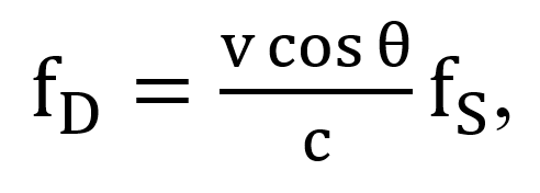
where fs is the Doppler shift frequency, v is the receiver (target) velocity, θ is the Doppler angle, and c is the speed of sound in tissue, which is 1540 m/s.
- Doppler shift frequency—The amount of change in frequency caused by the Doppler effect.
- dorsal—See posterior.
- duplex imaging—A process of combining two-dimensional real-time imaging with Doppler signal processing.
- echo—Within sonography, a reflected ultrasound signal. The signals are converted by the processor into gray dots on the ultrasound display.
- echocardiography—A diagnostic ultrasound test used to evaluate cardiac anatomy and physiology.
- eddies—Regions of circular flow patterns present within turbulent flow patterns.
- edema—Swelling caused by too much fluid trapped in the body’s tissues.
- effusion—An excessive accumulation of fluid in a region or a structure.
- emergency medicine—A medical specialty involving care for patients with acute illnesses or injuries requiring immediate medical attention.
- emphysema—A lung condition that causes shortness of breath due to damaged air sacs in the lungs.
- fascia—A thin casing of connective tissue that surrounds and holds every organ, blood vessel, bone, nerve fiber, and muscle in place.
- fibrosis—Scarring and hardening of tissues and organs.
- flexion—Bending of an arm or leg. It’s a physical position that decreases the angle between the bones of the limb at a joint.
- focal length—The distance from the scanning face of a transducer to the focus.
- focus—A narrow point in the cross section of the ultrasound beam. To improve the resolution at a given depth, the transducer must be focused in order to narrow the beam width. This is performed by varying the number of transmitting and receiving elements and delaying the signal once received.
- frame—A single image produced by one complete scan of the sound beam.
- frame rate—The number of frames of echo information stored each second.
- frequency—The number of cycles per second. In diagnostic ultrasound, it is usually demonstrated in megahertz.
- friction—Within sonography, the force that resists the relative motion between blood and the cardiovascular walls.
- gain—The degree of amplification of the returning echo.
- gate—A device that allows only echoes from selected depths to pass.
- gestation—The process or period of development inside the womb between conception and birth.
- grating—The process in which an input ultrasound beam splits into multiple beams after passing tissue, resulting in the formation of side lobe artifacts.
- grayscale—The range of brightness between white and black. The dark color indicates the total absence of reflected light, and the bright represents reflected light.
- gynecology—A specialty of the female reproductive system, including the diagnosis and treatment of conditions, disorders, and diseases.
- harmonics—Frequencies that are even and odd multiples of one another.
- hertz—Unit of frequency, one cycle per second.
- hydrocephalus—A neurological disorder caused by an abnormal buildup of cerebrospinal fluid in the ventricles or cavities deep within the brain.
- hydronephrosis—Swelling of a kidney due to urine failing to drain properly from the kidney to the bladder.
- hyperechoic—Areas on the image with more reflected echoes that appear brighter than the surrounding tissue.
- hypertension—High systolic and/or diastolic blood pressure.
- hypoechoic—Areas on the image with fewer reflected echoes that appear darker than the surrounding tissue.
- hysterectomy—A surgical removal of the uterus.
- impedance—The resistance to the propagation of ultrasound waves through tissues. It is determined by the density of the tissue and the sound propagation speed.
- inertia—Resistance to change in the state of either rest or motion or the tendency to keep moving in a straight line at a constant velocity.
- inferior—Away from the head; lower (for example, the foot is part of the inferior extremity).
- intensity—The power per unit area, usually expressed in milliwatts per square centimeter in sonographic discussions. It is the ultrasound power transferred per unit area.
- internal medicine—A branch of medicine that deals with long-term care involving common and complex illnesses such as the diagnosis and treatment of cancer, infections, and diseases affecting the heart, blood, kidneys, joints, and digestive, respiratory, and vascular systems in adolescents, adults, and elderly individuals.
- intima-media thickness analysis—An ultrasound measurement of the thickness of the two inner layers of the carotid arteries—the intima and media. Abnormal thickening of the arterial walls of the carotid arteries indicates a high risk for heart attack or stroke.
- intrarenal perfusion—Fluid injection into the renal blood vessels to or from the kidneys.
- laminar flow—Demonstration of fluid moving in an organized direction as if it were made up of layers that glide over one another.
- lateral—Away from the midline of the body (for example, the little toe is located at the lateral side of the foot).
- lateral resolution—Refers to reflectors that lie perpendicular to the axis of the ultrasound beam. The resolution is related to the beam width; that is, the wider the beam, the poorer the lateral resolution. An ultrasound beam is the narrowest at its focal length, and this is where the lateral resolution will be the best.
- lateral rotation—Lateral rotation of the upper limb at the shoulder or lower limb at the hip involves turning the anterior surface of the limb away from the midline of the body. Lateral rotation is also referred to as exterior rotation.
- linear array—An array made of rectangular elements arranged in a line.
- linear phased array—A linear array that operates by applying voltage pulses to all elements with small time differences to direct sound waves out in various directions.
- linear sequenced array—A linear array that operates by applying voltage pulses to groups of elements in a sequence.
- Lister’s tubercle—A bony prominence located at the distal end of the radius.
- lumen—An inner open space or cavity of a tubular structure, as of a blood vessel or an intestine.
- maternity—The period during and immediately after pregnancy.
- mechanical index—A value used to estimate the cavitation effect during exposure to ultrasound energy.
- medial—Relating to the midline of the body (for example, the middle toe is located at the medial side of the foot).
- medial rotation—Medial rotation of the upper limb at the shoulder or lower limb at the hip involves turning the anterior surface of the limb toward the midline of the body. Medial rotation is also referred to as internal rotation.
- mirroring—An artifact on a spectral display caused by an inability to separate the forward and reverse signal processing channels properly.
- morphology—The scientific study of the structure and form of animals and plants.
- Morrison’s pouch—Area between the liver and right kidney.
- motion mode—A one-dimensional position-versus-time ultrasound view of all changing or moving reflectors. This is used in echocardiography.
- obstetrics—The branch of medicine that deals with caring for and treating individuals during pregnancy, childbirth, and the postbirth period as well as the care of the newborn.
- oligohydramnios—A condition where there is reduced amniotic fluid around the baby during pregnancy.
- ophthalmology—A branch of medicine involving anatomy, physiology, and conditions of the eye.
- paracolic gutters—Areas between the colon and the abdominal wall.
- pediatrics—A specialty providing medical care to infants, children, and adolescents.
- perinatologist—An obstetrician-gynecologist who specializes in high-risk pregnancies.
- period—Time per cycle.
- physiatry—A branch of medicine that deals with the treatment of injuries or illnesses of the nerves, muscles, and bones. Also known as physical medicine and rehabilitation.
- piezoelectric effect—The ability of certain crystals to generate an electric charge in response to applied mechanical stress. Piezoelectric materials produce electrical signals when deformed or vice versa (i.e., the crystal changes shape and vibrates when a voltage is applied to it). These crystals are the primary components of ultrasound transducers.
- pixel—Picture elements; the unit into which imaging information is divided for storage and display.
- polyhydramnios—A condition when there is too much amniotic fluid around the baby during pregnancy.
- posterior—Relating to the back (for example, the shoulder blades are located on the posterior side of the body).
- potential or power of hydrogen—A measure of hydrogen ion concentration representing the acidity or basicity of aqueous or other liquid solutions.
- pouch of Douglas—The caudal extension of the peritoneal cavity. It is the rectovaginal pouch in the female and the rectovesical pouch in the male.
- propagation speed—The speed at which a wave travels through a medium and is equal to the product of the wavelength and frequency. The average propagation speed of sound waves in a soft tissue is 1540 meters/second.
- proximal—Toward or nearest the trunk or the point of origin of a part (for example, the proximal end of the femur joins with the pelvic bone).
- pulsatility index—A dimensionless parameter that is used to assess pulsatility and is defined as the difference between maximum and minimum blood flow velocity, normalized to the average velocity.
- pulsed Doppler—A Doppler mechanism that transmits a coherent burst of sound waves into the tissue and waits for returning echoes from a specific location, then processes these echoes for Doppler shift frequency content.
- pulse repetition frequency—The number of pulses per second.
- pulse repetition period—Interval of time from the beginning of one pulse to the beginning of the next.
- rarefaction—A region in a longitudinal wave (like sound waves) where the particles are farthest apart.
- real-time display—Immediate display of an ultrasound scan of moving structures made possible by the use of higher frame rates.
- reflection—Portion of the sound returned from a medial boundary (i.e., refers to the bending of the ultrasound beam at the boundary surface between two mediums with different acoustic impedances).
- refraction—Change of sound direction on passing from one medium to another.
- regurgitation—The flow of a fluid through a vessel or valve in the body in a direction opposite to normal.
- renal arterial resistive index—An index used to assess the risk for renal arterial disease. It is commonly used to indicate renal function, pathology, prognosis, and responsiveness to steroid therapy in chronic kidney disease patients.
- resolution—Within sonography, represents the ability of an ultrasound system to distinguish between two closely spaced reflectors as separate structures.
- reverberation—Artifact resulting from false echoes produced due to repeated reflections between two interfaces with a high acoustic impedance mismatch.
- sagittal plane or view—An anatomical plane that divides the body into right and left sections.
- scattering—Spreading of ultrasound beam in different directions as it strikes a boundary or interface between two small structures.
- shadowing—Reduction in echo amplitude from reflectors behind a strongly reflecting or attenuating structure.
- sonography—The process of visualizing body structures using ultrasound.
- spatial—Relating to the position, area, and size of things.
- spatial resolution—The minimum distance between two adjacent features that can be detected by the imaging system.
- spatiotemporal—Relating to both space and time.
- spectral analysis—Separation of frequencies in a Doppler signal for display as a Doppler spectrum in the form of a neat graph.
- specular reflection—An optical process involving reflections from a large, smooth boundary.
- stenosis—An abnormal narrowing of a blood vessel or other tubular organ or structure such as foramina and canals.
- superior—Toward the head end of the body; upper (for example, the hand is part of the superior extremity).
- supine—A position in which a person lies on their back with their face and torso pointing up.
- surgery—An invasive use of instruments to test, remove, or adjust an individual’s original anatomy for treatment of illness, injury, or disease or cosmetic purposes.
- systemic hemodynamics—Study of the relationship between arterial blood pressure and blood flow. Systemic hemodynamic state is the mean value of blood pressure and the mean value of blood flow over one heartbeat interval.
- systole—Phase of the cardiac cycle in which the heart contracts and pumps out the blood.
- temporal—Relating to time.
- thermal index—A value used as an indicator for the potential risk of tissue heating due to exposure to ultrasound.
- thrombosis—The development of blood clots inside blood vessels that obstruct blood flow in the circulatory system.
- thrombus—A blood clot in the circulatory system.
- transducer—A device that converts variations in acoustic pressure to an electrical signal or vice versa.
- transmitter—A device that uses an electrical current to generate acoustic vibrations or pulses.
- transverse plane or view—An anatomical plane that divides the body into superior and inferior sections.
- trimester—A period of three months during gestation. The human gestation period has three trimesters (first, second, and third).
- turbulent flow—A nonlaminar flow condition in which the kinetic energy of flow creates vortices. The overall fluid motion is complex and chaotic.
- urology—The study or medical treatment of diseases and conditions of the urinary tract system and certain reproductive organs.
- valves—A flap or leaflet, usually inside a tubular structure, that acts as a one-way inlet and permits fluids or gases to flow in just one direction.
- ventilator—A device that supports or re-creates the process of breathing by pumping air into the lungs when a person is not able to breathe enough on their own.
- ventral—See anterior.
- viscosity—A measure of the resistance of a fluid to flow. It is an indicator of the internal friction of a moving fluid. For example, a fluid with high viscosity flows slower than one with low viscosity.
