
Attributions
Chapter 1
Figure 1.1 – Clay model of a sheep’s liver, Babylonian Temple School © Wellcome Images,Wellcome Images Library, London is licensed under a CC BY (Attribution) license
Figure 1.2 – The Edwin Smith Papyrus © Edited by Jeff Dahl is licensed under a CC0 (Creative Commons Zero) license
Figure 1.3 – Huang di nei jing su wen © Bing Wang, 1115, Library of Congress is licensed under a CC0 (Creative Commons Zero) license
Figure 1.4 – Susrutea-Samhita, 12-13th Century © Los Angeles County Museum of Art is licensed under a CC0 (Creative Commons Zero) license
Figure 1.5 – L0020565 Galen, Opera omnia, dissection of a pig. © Wellcome Library, London. Wellcome Images images@wellcome.ac.uk is licensed under a CC BY (Attribution) license
Figure 1.6 – Photograph of statue of Ibn Al Nafis © Takrouri & Khalaf 2003 is licensed under a CC BY (Attribution) license
Figure 1.7 – L0031072 A. Vesalius, De humani corporis fabrica libri septum © Wellcome Library, London. Wellcome Images images@wellcome.ac.uk is licensed under a CC BY (Attribution) license
Figure 1.8 – L’Histoire de la nature des oyseaux, Belon, 1555 © Gallica Digital Library and is available under the digital ID btv1b8608302w/f74 is licensed under a CC0 (Creative Commons Zero) license
Figure 1.9 – Elementorum myologiae specimen, Neils Steensen, 1667 © Available in the BEIC digital library and uploaded in partnership with BEIC Foundation. is licensed under a CC0 (Creative Commons Zero) license
Figure 1.10 – Ear, wax model, Anna Morandi Manzolini © Photo taken by Warburg. is licensed under a CC BY (Attribution) license
Figure 1.11 – V0018739 William Hunter: his home and anatomy school, London © Wellcome Library, London. Wellcome Images is licensed under a CC BY (Attribution) license
Figure 1.12 – Illustration of resurrectionists at work, accompanying the story of John Holmes and Peter Williams, whipped for stealing dead bodies., Habolt Knight Browne, 1887. © https://archive.org/stream/chroniclesofcri01pelh#page/n317/mode/2up is licensed under a CC0 (Creative Commons Zero) license
Figure 1.13 – Histore naturelle, vol. 10, Georges-Louis Leclerc de Buffon, 1763 © Part of Biblioteca Europea di Informazione e Cultura is licensed under a CC0 (Creative Commons Zero) license
Figure 1.14 – Lamarckian evolution, 2009, Solarist. is licensed under a CC BY (Attribution) license
Figure 1.15 – Plate 73, “Le règne animal distribué d’après son organisation” , Georges Cuvier, vol. 8, second edition, 1828. © Georges Cuvier, vol. 8, second edition, 1828. is licensed under a CC BY (Attribution) license
Figure 1.16 – Charles Darwin & Alfred Wallace © OpenStax 2e is licensed under a CC BY (Attribution) license
Figure 1.17 – Drawing of skull © Thomas Henry HuxleyCharles Joseph Singer is licensed under a CC BY (Attribution) license
Chapter 2
Figure 2.1 – A general phylogenetic (evolutionary) tree of animals © Lisa B. Whitenack is licensed under a CC BY (Attribution) license
Figure 2.2 – Figure 29.3 Chordate features. In chordates, four common features appear at some point during development: a notochord, a dorsal hollow nerve cord, pharyngeal slits, and a post-anal tail. The endostyle is embedded in the floor of the pharynx. © OpenStax Biology 2e is licensed under a CC BY (Attribution) license
Figure 2.3 – Anatomical directions and planes of section on a kangaroo © Jmerritt at English Wikipedia is licensed under a CC BY (Attribution) license
Figure 2.4 – Medical gallery of Blausen Medical 2014 © Blausen.com staff, WikiJournal of Medicine 1 (2) is licensed under a CC BY (Attribution) license
Figure 2.5 – Regions of the Human Body © OpenStax Biology 2e is licensed under a CC BY (Attribution) license
Figure 2.6 – A generalized tree of the vertebrates with tree anatomy labeled © Lisa B. Whitenack is licensed under a CC BY (Attribution) license
Figure 2.7 – Protostome deueterostome development © Yassine Mrabet is licensed under a CC BY (Attribution) license
Figure 2.8 – Echinoderm diversity is licensed under a Public Domain license
Figure 2.9 – Echinoderm larva © Bruno C. Vellutini is licensed under a CC BY (Attribution) license
Figure 2.10 – Pterobranchia representatives © https://collections.nmnh.si.edu/search/iz/?ark=ark:/65665/3db6c2545c10b444189b382fd11ec9e96 AND Encyclopædia Britannica (11th ed.), v. 22, 1911, “Pterobranchia,” fig. 1, p. 614. is licensed under a CC0 (Creative Commons Zero) license
Figure 2.11 – Enteropneusts © Spengel, Johann Wilhelm (1893) Die Enteropneusten des Golfes von Neapel und der angrenzenden Meeres-Abschnitte Berlin : Verlag von R. Friedländer & Sohn is licensed under a Public Domain license
Figure 2.12 – Dorsoventral Patterning in Hemichordates: Insights into Early Chordate Evolution. © Lowe CJ, Terasaki M, Wu M, Freeman RM Jr, Runft L, et al is licensed under a CC BY (Attribution) license
Figure 2.13 – Larval Forms © “Encyclopædia Britannica (11th ed.), v. 16, 1911, “Larval Forms,” p. 226, fig. 5. AND Bruno C. Vellutini” is licensed under a CC0 (Creative Commons Zero) license
Figure 2.14 – Chordate Diversity © User:Biopics User:Nhobgood User:Albert kok User:Fir0002 is licensed under a CC BY (Attribution) license
Figure 2.15 – Urochordate Diversity © Rickard Zerpe AND Lars Plougmann from London, United Kingdom is licensed under a CC BY (Attribution) license
Figure 2.16 – Tunicate Anatomy © OpenStax is licensed under a CC BY (Attribution) license
Figure 2.17 – Cephalochordate anatomy © Systematicist AND Encyclopædia Britannica Eleventh Edition, Vol. 1, Page 887 is licensed under a CC BY (Attribution) license
Figure 2.18 – Chordata and Ambulacraria phylogeny © Lisa B. Whitenack is licensed under a CC BY (Attribution) license
Figure 2.19 – Early Vertebrates © Degan Shu, Northwest University, Xi’an, China is licensed under a CC BY (Attribution) license
Chapter 3
Figure 3.1 – Neural Crest © NikNaks is licensed under a Public Domain license
Figure 3.2 – The cranial sensory nervous system: specification of sensory progenitors and placodes © Streit, A., (December 15, 2008), StemBook, ed. The Stem Cell Research Community, StemBook, doi/10.3824/stembook.1.31.1, http://www.stembook.org. is licensed under a CC BY (Attribution) license
Figure 3.3 – Embryonic brain top © Nrets, vector conversion by Surachit is licensed under a CC BY (Attribution) license
Figure 3.4 – Thoracic Vertebra © Lisa B. Whitenack is licensed under a CC BY (Attribution) license
Figure 3.5 – Hox Genes: Master Regulators of the Animal Bodyplan © A.J. Durston is licensed under a CC BY (Attribution) license
Figure 3.6 – Phylogenetic Tree of the Vertebrata © Lisa B. Whitenack is licensed under a CC BY (Attribution) license
Figure 3.7 – Eptatretus deani © Bulletin of the United States Fish Commission 1906 is licensed under a Public Domain license
Figure 3.8 – Petromyzon marinus © Fernando Losada Rodríguez is licensed under a CC BY (Attribution) license
Figure 3.9 – Hagfish knot © JLplusAL is licensed under a CC BY (Attribution) license
Figure 3.10 – Hagfish Slime as a Defense Mechanism against Gill-breathing Predators © Zintzen, Vincent, Clive D. Roberts, Marti J. Anderson, Andrew L. Stewart, Carl D. Struthers, and Euan S. Harvey. is licensed under a CC BY (Attribution) license
Figure 3.11 – Tiszai ingola balinon © Zsoldos Márton is licensed under a CC BY (Attribution) license
Figure 3.12 – The geological time scale for the Phanerozoic Eon © Lisa B. Whitenack is licensed under a CC BY (Attribution) license
Figure 3.13 – Geologic Clock with events and periods © “Woudloper Derivative work: Hardwigg is licensed under a Public Domain license
Figure 3.14 – Conodonta © Magdaléna Kostková is licensed under a Public Domain license
Figure 3.15 – Iowagnathus grandus © Derek E.G. Briggs; Huaibao P. Liu; Robert M. McKay; Brian J. Witzke is licensed under a CC BY (Attribution) license
Figure 3.16 – Hindeodus elements © Muhui Zhang, Haishui Jiang, Mark A. Purnell, Xulong Lai is licensed under a CC BY (Attribution) license
Figure 3.17 – Fossil Fish © Jon Houseman is licensed under a CC BY (Attribution) license
Figure 3.18 – Dunkleosteus Sannoble © Mitternacht90 is licensed under a Public Domain license
Figure 3.19 – Dunkleosteus terrelli size © PaleoNeolitic is licensed under a CC0 (Creative Commons Zero) license
Figure 3.20 – Bothriolepis canadensis based on 2014 reconstruction © Paleobiome is licensed under a CC0 (Creative Commons Zero) license
Figure 3.21 – Chondrichtyes composite © Albert kok, Columbus zoo is licensed under a CC BY (Attribution) license
Figure 3.22 – Extinct chondrichthyes © Géry PARENT, James St. John, Stanton F. Fink is licensed under a CC BY (Attribution) license
Figure 3.23 – Chondrichthyes cartilage structure © Seidel, R., Jayasankar, A.K., Dean, M.N. (2020): The multiscale architecture of tessellated cartilage and its relation to function. Jornal of Fish Biology (DOI: 10.1111/jfb.14444) is licensed under a CC BY (Attribution) license
Figure 3.24 – Diplacanthus striatus fossil fish © James St. John is licensed under a CC BY (Attribution) license
Figure 3.25 – Acanthodi NT © Nobu Tamura Email:nobu.tamura@yahoo.com is licensed under a CC BY (Attribution) license
Figure 3.26 – Actinopts © MathKnight made the composite figure is licensed under a CC BY (Attribution) license
Figure 3.27 – Epinephelus merra JNC3054 Pectoral fin on white background © Jean-Lou Justine is licensed under a CC BY (Attribution) license
Figure 3.28 – Polypterus senegalus (male and female) © TVRGolf is licensed under a CC BY (Attribution) license
Figure 3.29 – Paddlefish © U.S. Fish and Wildlife Service is licensed under a Public Domain license
Figure 3.30 – Amia calva © Stan Shebs is licensed under a CC BY (Attribution) license
Figure 3.31 – Micropterus salmoides captown © Nervurax Fishing is licensed under a CC0 (Creative Commons Zero) license
Figure 3.32 – Toothed Oral and Pharyngeal Jaws © Gareth J Fraser is licensed under a CC BY-SA (Attribution ShareAlike) license
Figure 3.33 – Coelocanth drawing © No machine-readable author provided. Bogdan assumed (based on copyright claims). is licensed under a CC BY (Attribution) license
Figure 3.34 – Sarcopterygii © “User:ArthurWeasley User:Bruce A.S.Henderson User:Mitch Ames User:DiBgd” is licensed under a CC BY-SA (Attribution ShareAlike) license
Figure 3.35 – Marjorie Courtenay-Latimer and Coelacanth © A Christmas Story: 22 December 1938. The South African Institute for Aquatic Biodiversity. is licensed under a Public Domain license
Figure 3.36 – Marbled lungfish © http://opencage.info/pics.e/large_11454.asp is licensed under a CC BY (Attribution) license
Figure 3.37 – Tiktaalik Chicago © Eduard Solà is licensed under a CC BY-SA (Attribution ShareAlike) license
Figure 3.38 – Tiktaalik roseae life restor © Zina Deretsky, National Science Foundation is licensed under a Public Domain license
Figure 3.39 – A phylogenetic tree of the Tetrapodomorpha © Lisa B. Whitenack is licensed under a CC BY (Attribution) license
Figure 3.40 – Simplified phylogeny spanning the fish–tetrapod transition © Per E. AHLBERG is licensed under a CC BY (Attribution) license
Figure 3.41 – Temnospondyl skulls © Daderot and Emőke Dénes is licensed under a CC BY (Attribution) license
Figure 3.42 – The temnospondyl Eryops © Camelops is licensed under a CC BY (Attribution) license
Figure 3.43 – Diplocaulus © Camelops is licensed under a CC BY (Attribution) license
Figure 3.44 – Brachydectes © Smokeybjb is licensed under a CC BY (Attribution) license
Figure 3.45 – Amphibians © “http://calphotos.berkeley.edu Author Franco Andreone – see author; https://www.inaturalist.org/photos/102423969ization; AND LiquidGhoul” is licensed under a CC BY (Attribution) license
Figure 3.46 – Reconstructions of nonamniote reptilomorphs: Archeria2DB and Seymouria2 © Dmitry Bogdanov is licensed under a CC BY-SA (Attribution ShareAlike) license
Figure 3.47 – Dimetrodon skull lateral © Smokeybjb is licensed under a CC BY-SA (Attribution ShareAlike) license
Figure 3.48 – Various pelycosaurs © Nobu Tamura (http://spinops.blogspot.com) AND Jonathan Chen AND DiBgd AND Smokeybjb is licensed under a CC BY-SA (Attribution ShareAlike) license
Figure 3.49 – Therapsida © Prehistoricplanes is licensed under a CC BY-SA (Attribution ShareAlike) license
Figure 3.50 – Scymnognathus © Eden, Janine and Jim (https://www.flickr.com/photos/edenpictures/42384543145/) is licensed under a CC BY-SA (Attribution ShareAlike) license
Figure 3.51 – Cynodont © Lutz Benseler is licensed under a CC BY-SA (Attribution ShareAlike) license
Figure 3.52 – Monotreme collage © Ypna is licensed under a CC BY-SA (Attribution ShareAlike) license
Figure 3.53 – Marsupialia © LittleJerry is licensed under a CC BY-SA (Attribution ShareAlike) license
Figure 3.54 – Placental Mammals 2021 Montage © The0Quester is licensed under a Public Domain license
Figure 3.55 – Parareptilia © “Jonathan Chen AND Dmitry Bogdanov wikidata:Q39957193 AND Nobu Tamura (http://spinops.blogspot.com)” is licensed under a CC BY-SA (Attribution ShareAlike) license
Figure 3.56 – Rhynchocephalia © Tiouraren (Y.-C. Tsai) AND KeresH is licensed under a CC BY-SA (Attribution ShareAlike) license
Figure 3.57 – Squamata © “File:Anelytropsis papillosus, Mexican Blind Lizard, Tamaulipas.jpg: William L. Farr File:Uroplatus fimbriatus (3).jpg: TimVickers File:BennyTrapp Chamaeleo chamaeleon Samos Griechenland.jpg: Benny Trapp File:Lacerta agilis male 2.JPG: Christoph Caina File:White-throated Monitor (Varanus albigularis) looking for food … (30427772662).jpg: Bernard DUPONT from FRANCE File:2017.07.17.-17-Tiefer See oder Grubensee-Storkow (Mark)–Ringelnatter.jpg: Andreas Eichler File:Euspondylus chasqui, male.jpg: Germán Chávez, Karen Siu-Ting, Vilma Duran, Pablo J. Venegas File:Amphisbaena alba03.jpg: Diogo B. Provete File:Blue-toungued skink444.jpg: Benjamint444” is licensed under a CC BY-SA (Attribution ShareAlike) license
Figure 3.58 – The skeleton and a reconstruction of the mosasaur Halisaurus © “Source Ybmos_1b Uploaded by FunkMonk Author Greg Goebel from Loveland CO, USA AND Nobu Tamura (http://spinops.blogspot.com)” is licensed under a CC BY (Attribution) license
Figure 3.59 – Extant Testudines © “File:Rotbauchspitzkopfschildkroete-07.jpg File:Indian flapshell turtle with Spots visible 03.jpg File:Galapagos giant tortoise Geochelone elephantopus.jpg File:Hawksbill Turtle.jpg” is licensed under a CC BY-SA (Attribution ShareAlike) license
Figure 3.60 – Turtle anatomy © “English: Comparative zoology, structural and systematic : for use in schools and colleges 1883, New York : Harper & Bros.” is licensed under a CC0 (Creative Commons Zero) license
Figure 3.61 – Archosauria © MathKnight is licensed under a CC BY-SA (Attribution ShareAlike) license
Figure 3.62 – Archosaur skull © Jcwf is licensed under a CC0 (Creative Commons Zero) license
Figure 3.63 – Phylogenetic framework © “Lee et al. 2020. Evolutionary and ontogenetic changes of the anatomical organization and modularity in the skull of archosaurs. Scientific Reports :16138 |” is licensed under a CC BY-SA (Attribution ShareAlike) license
Figure 3.64 – Various pseudosuchians © “Postosuchus kirkpatricki Uploaded by FunkMonk AND Saurosuchus_BW.jpg: ArthurWeasley aweasley@hotmail.com Two Gharials.jpg: Harsha K R from Bangalore, India Angistorhinus_BW.jpg (only included in 1st version, April 2009): Nobu Tamura Longosuchus_BW.jpg: ArthurWeasley aweasley@hotmail.com Chenanisuchus_BW.jpg: ArthurWeasley Dakosaurus2.jpg: Dmitry Bogdanov Litargosuchus leptorhynchus.jpg: Smokeybjb derivative work: Matt Martyniuk (talk) ” is licensed under a CC BY-SA (Attribution ShareAlike) license
Figure 3.65 – A typical crurotarsal ankle found in Pseudosuchia and a typical mesotarsal ankle found in Avemetatarsalia © UC Museum of Paleontology, www.ucmp.berkeley.edu is licensed under a CC BY-NC-SA (Attribution NonCommercial ShareAlike) license
Figure 3.66 – Examples of Pterosauria © “Dmitry Bogdanov wikidata:Q39957193 AND Tim Evanson AND Armin Reindl AND Slate Weasel” is licensed under a CC BY-SA (Attribution ShareAlike) license
Figure 3.67 – Dinosaur hip structure. Saurischia pelvis structure AND Ornithischia pelvis structure © Fred the Oyster is licensed under a CC BY-SA (Attribution ShareAlike) license
Figure 3.68 – Ornithischia montage © “Heterodontosaurus_tucki_cast_-_University_of_California_Museum_of_Paleontology_-_Berkeley,_CA_-_DSC04696.JPG Nipponosaurus.jpg Stegosaurus_ungulatus.jpg Borealopelta_Royal_Tyrrell_2.jpg Royal_Tyrrell_Museum_Stegoceras.jpg Triceratops_Specimen_at_the_Houston_Museum_of_Natural_Science_v01.jpg Author User:Daderot [1] [2] User:Etemenaki3 [3] User:Agsftw” is licensed under a CC BY-SA (Attribution ShareAlike) license
Figure 3.69 – Sauropodomorpha © “Fanboyphilosopher (File:Eoraptor Deep Time.jpg) FunkMonk (File:Plateosaurus Skelett 2.jpg) Ad Meskens (File:Brontosaurus Yale Peabody cropped.jpg) Ghedoghedo (File:Europasaurus holgeri.jpg) James St. John (File:Mamenchisaurus hochuanensis Field Museum.jpg) Kabacchi (File:Nigersaurus mount.jpg) William Irvin Sellers, Lee Margetts, Rodolfo Aníbal Coria, Phillip Lars Manning (File:Argentinosaurus skeleton, PLoS ONE.png) ScottRobertAnselmo (File:CM Diplodocus.jpg) AStrangerintheAlps (File:FMNH Brachiosaurus.JPG)” is licensed under a CC BY-SA (Attribution ShareAlike) license
Figure 3.70 – Theropod dinosaurs © “This file was derived from: Allosaurus_SDNHM.jpg: Sheep81 AND Jens Lallensack AND Dinoguy2 ” is licensed under a CC BY-SA (Attribution ShareAlike) license
Figure 3.71 – Three equally plausible phylogenetic trees © Lisa B. Whitenack is licensed under a CC BY (Attribution) license
Figure 3.72 – Examples of paravian dinosaurs © James St. John, Ohio State University, Newark AND NobuTamura http://paleoexhibit.blogspot.com/ http://spinops.blogspot.com/ AND Emily Willoughby (emily.a.willoughby@gmail.com) is licensed under a CC BY-SA (Attribution ShareAlike) license
Figure 3.73 – Birds and fossils © James St. John, Ohio State University, Newark AND NobuTamura http://paleoexhibit.blogspot.com/ http://spinops.blogspot.com/ AND Emily Willoughby (emily.a.willoughby@gmail.com) is licensed under a CC BY-SA (Attribution ShareAlike) license
Figure 3.74 – Bird Diversity 2013 © “ade from the following Wikimedia free images (some cropped and/or rotated): Row 1 (left to right) Red-crested Turaco RWD.jpg — Red-crested Turaco (Tauraco erythrolophus) Balaeniceps rex – Weltvogelpark Walsrode 09-2010 2.jpg — Shoebill (Balaeniceps rex) Phaethon lepturus (Warwick, Bermuda).jpg — White-tailed Tropicbird (Phaethon lepturus) Row 2 Haliaeetus pelagicus (Rausu, Japan).jpg — Steller’s Sea Eagle (Haliaeetus pelagicus) Balearica regulorum (Tarime, Tanzania).jpg — Grey Crowned Crane (Balearica regulorum) Pavo cristatus -Castellar Zoo, Castellar de la Frontera, Spain-8a.png — Indian Peafowl (Pavo cristatus) Row 3 File:Rock dove – natures pics.jpg — Rock Dove(Columba livia) Calypte anna -San Luis Obispo, California, USA -male -flying-8.jpg — Anna’s Hummingbird (Calypte anna) Puffin Latrabjarg Iceland.jpg — Atlantic Puffin (Fratercula arctica) Row 4 Casuarius casuarius (Carmoo QLD, Australia).jpg — Southern Cassowary (Casuarius casuarius) Trichoglossus haematodus (Melbourne, Australia).jpg — Rainbow Lorikeet (Trichoglossus haematodus moluccanus) Caribbean flamingo at slimbridge arp.jpg — American Flamingo (Phoenicopterus ruber) Row 5 Antarctic, gentoo penguin (js) 16.jpg — Gentoo Penguin (Pygoscelis papua) Ardea cinerea 1.JPG — Grey Heron (Ardea cinerea) Sula nebouxii -Santa Cruz, Galapagos Islands, Ecuador-8 (2).jpg — Blue-footed Booby (Sula nebouxii) Row 6 Minla strigula – Doi Inthanon.jpg — Bar-throated Minla (Minla strigula) Eurasian Eagle-Owl RWD.jpg — Eurasian Eagle-Owl (Bubo bubo) Keel-billed toucan woodland.jpg — Keel-billed Toucan (Ramphastos sulfuratus)” is licensed under a CC BY-SA (Attribution ShareAlike) license
Figure 3.75 – Aepyornis and Dromornis stirtoni © Kevmin AND Museon, Den Haag is licensed under a CC BY-SA (Attribution ShareAlike) license
Chapter 4
Figure 4.1 – Final stem cell differentiation (1) © Haileyfournier is licensed under a CC BY-SA (Attribution ShareAlike) license
Figure 4.2 – Auge Entwicklung © P. Stanka is licensed under a CC BY-SA (Attribution ShareAlike) license
Figure 4.3 – Deer sperm © Ros-Santaella JL, Pintus E, Garde JJ. 2015. Intramale variation in sperm size: functional significance in a polygynous mammal. PeerJ 3:e1478 is licensed under a CC BY (Attribution) license
Figure 4.4 – Macrolecithal zebrafish embryo and Human oocyte © Swinburne et al., 2011, PLoS ONE is licensed under a CC BY (Attribution) license
Figure 4.5 – Ovocyte II © Samuel J. Holmes; Kuroda et al.; Zachary Wilson; NinaSes; Swift D; Hill, M.A. is licensed under a CC BY-SA (Attribution ShareAlike) license
Figure 4.6 – Diagram of Blastocyst stage © Zachary Wilson / CK-12 Foundation is licensed under a CC BY-SA (Attribution ShareAlike) license
Figure 4.7 – Gastrulation in a chicken embryo © Serrano Nájera G, Weijer CJ. The evolution of gastrulation morphologies. Development. 2023 Apr 1;150(7):dev200885. doi: 10.1242/dev.200885. is licensed under a CC BY (Attribution) license
Figure 4.8 – Derivatives of the mammalian inner cell mass © Shparberg RA, Glover HJ and Morris MB (2019) Modeling Mammalian Commitment to the Neural Lineage Using Embryos and Embryonic Stem Cells. Front. Physiol. 10:705. doi: 10.3389/fphys.2019.00705 is licensed under a CC BY (Attribution) license
Figure 4.9 – Neural crest © NikNaks is licensed under a Public Domain license
Figure 4.10 – Mesoderm regions and their major derivatives © Tani, S., Chung, Ui., Ohba, S. et al. Understanding paraxial mesoderm development and sclerotome specification for skeletal repair. Exp Mol Med 52, 1166–1177 (2020). https://doi.org/10.1038/s12276-020-0482-1 is licensed under a CC BY (Attribution) license
Figure 4.11 – Microscopy views © Hester SD, Belmonte JM, Gens JS, Clendenon SG, Glazier JA (2011) A Multi-cell, Multi-scale Model of Vertebrate Segmentation and Somite Formation. PLoS Comput Biol 7(10): e1002155. https://doi.org/10.1371/journal.pcbi.1002155 is licensed under a CC BY (Attribution) license
Figure 4.12 – Diagram of mouse embryo © Thawani A and Groves AK (2020) Building the Border: Development of the Chordate Neural Plate Border Region and Its Derivatives. Front. Physiol. 11:608880. doi: 10.3389/fphys.2020.608880 is licensed under a CC BY (Attribution) license
Figure 4.13 – Embryonic brain © Nrets, vector conversion by Surachit is licensed under a CC BY-SA (Attribution ShareAlike) license
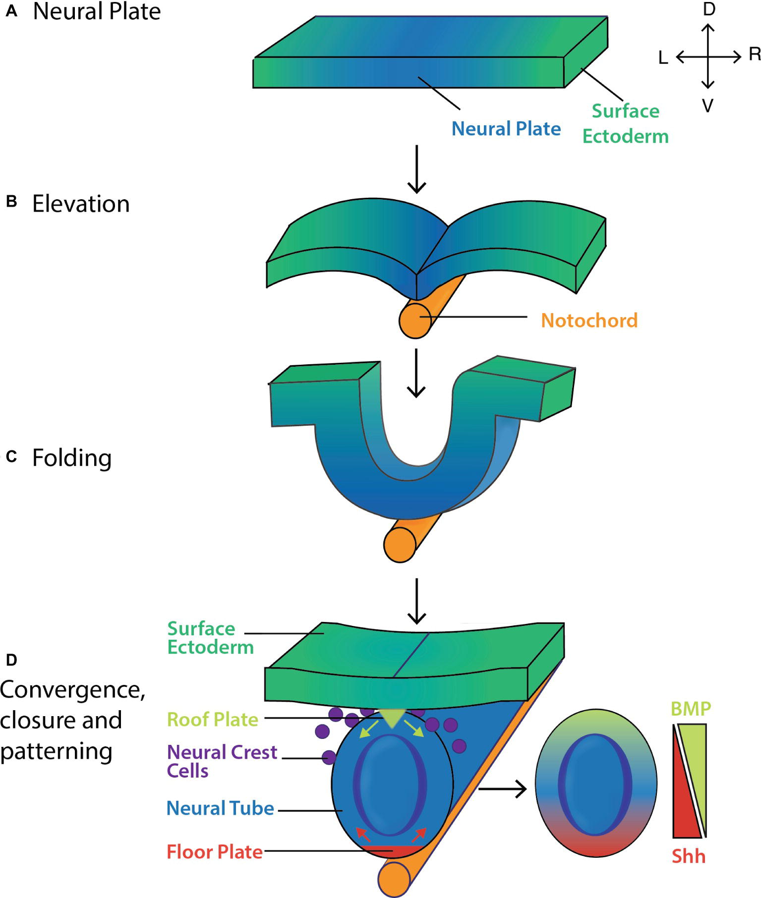
Figure 4.14 – Neural tube signaling © Thawani A and Groves AK (2020) Building the Border: Development of the Chordate Neural Plate Border Region and Its Derivatives. Front. Physiol. 11:608880. doi: 10.3389/fphys.2020.608880 is licensed under a CC BY (Attribution) license
Figure 4.15 – Chem. Synapse scheme is licensed under a Public Domain license
Figure 4.16 – Hox genes © Durston, A. J. (2012). Hox Genes: Master Regulators of the Animal Bodyplan. InTech. doi: 10.5772/37007 is licensed under a CC BY-SA (Attribution ShareAlike) license
Figure 4.17 – Hox in rhombomeres © Trainor, Paul A., Making Headway: The Roles of Hox Genes and Neural Crest Cells in Craniofacial Development, The Scientific World Journal, 3, 967108, 25 pages, 2003. https://doi.org/10.1100/tsw.2003.11 is licensed under a CC BY (Attribution) license
Figure 4.18 – Stylopod-zygopod-autopod © Peteruetz is licensed under a CC BY-SA (Attribution ShareAlike) license
Figure 4.19 – Limb bud diagram © Sisi Chen is licensed under a CC BY-SA (Attribution ShareAlike) license
Figure 4.20 – Gene expression bat wing © Zebra.element is licensed under a Public Domain license
Figure 4.21 – Webbed feet phylogeny © Tokita, M., Matsushita, H. & Asakura, Y. Developmental mechanisms underlying webbed foot morphological diversity in waterbirds. Sci Rep 10, 8028 (2020). https://doi.org/10.1038/s41598-020-64786-8 is licensed under a CC BY (Attribution) license
Figure 4.22 – Organizer experiment © VAN Robays J. Hilde Mangold-Pröscholdt (1898 – 1924): The Spemann-Mangold Organizer. Facts Views Vis Obgyn. 2016 Mar 28;8(1):63-68. is licensed under a CC BY (Attribution) license
Chapter 5
Figure 5.1 – Biomaterials © Kelly M. Diamond and Vanessa K Hilliard is licensed under a CC BY (Attribution) license
Figure 5.2 – Units © Kelly M. Diamond and Vanessa K Hilliard is licensed under a CC BY (Attribution) license
Figure 5.3 – Forces © Kelly M. Diamond and Vanessa K Hilliard is licensed under a CC BY (Attribution) license
Figure 5.4 – Scaling © Kelly M. Diamond and Vanessa K Hilliard is licensed under a CC BY (Attribution) license
Figure 5.5 – Surface area to volume ratios in the wild © Denali National Park and Reserve; Joshua Tree National Park is licensed under a Public Domain license
Figure 5.6 – Monsters © Kelly M. Diamond and Vanessa K Hilliard is licensed under a CC BY (Attribution) license
Figure 5.7 – Stress-Strain © Kelly M. Diamond and Vanessa K Hilliard is licensed under a CC BY (Attribution) license
Figure 5.8 – Lever systems © Kelly M. Diamond and Vanessa K Hilliard is licensed under a CC BY (Attribution) license
Figure 5.9 – Loading regimes © Kelly M. Diamond and Vanessa K Hilliard is licensed under a CC BY (Attribution) license
Figure 5.10 – Stress-strain © Kelly M. Diamond and Vanessa K Hilliard is licensed under a CC BY (Attribution) license
Figure 5.11 – Drag © Kelly M. Diamond and Vanessa K Hilliard is licensed under a CC BY (Attribution) license
Figure 5.12 – Lift © Kelly M. Diamond and Vanessa K Hilliard is licensed under a CC BY (Attribution) license
Figure 5.13 – Posture © Kelly M. Diamond and Vanessa K Hilliard is licensed under a CC BY (Attribution) license
Figure 5.14 – Form-function © Kelly M. Diamond and Vanessa K Hilliard is licensed under a CC BY (Attribution) license
Figure 5.15 – Feeding morphology © Kelly M. Diamond and Vanessa K Hilliard is licensed under a CC BY (Attribution) license
Chapter 6
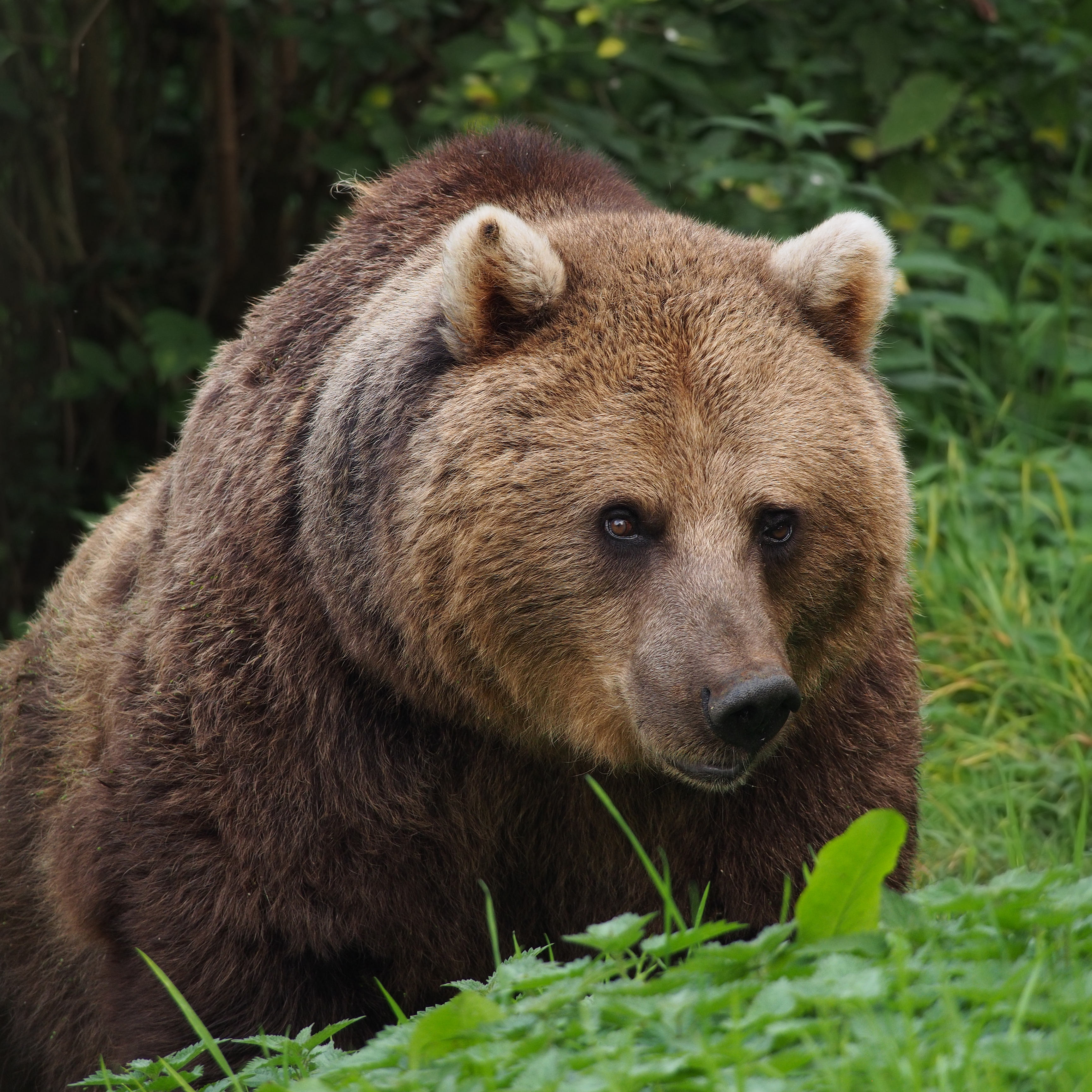
Figure 6.1 – Integument Derivatives © European Brown Bear, Francis Franklin; Goldfish scales, Maky Orel; Flaco, David Barrett; Moose, Ryan Hagerty; Komodo dragon claws, Charles Sharp is licensed under a CC BY-SA (Attribution ShareAlike) license
Figure 6.2 – Human 01 © Laboratoires Servier is licensed under a CC BY-SA (Attribution ShareAlike) license
Figure 6.3 – Shark collagen © Bill Ryerson is licensed under a CC BY (Attribution) license
Figure 6.4 – Dunkleosteus © Engelman, R.K. A (2023). Devonian Fish Tale: A New Method of Body Length Estimation Suggests Much Smaller Sizes for Dunkleosteus terrelli (Placodermi: Arthrodira). Diversity 2023, 15, 318. https://doi.org/10.3390/d15030318 is licensed under a CC BY-SA (Attribution ShareAlike) license
Figure 6.5 – Denticles © Viana STFL, Carvalho MR (2020) Squalus shiraii sp. nov. (Squaliformes, Squalidae), a new species of dogfish shark from Japan with regional nominal species revisited. Zoosystematics and Evolution 96(2): 275-311. https://doi.org/10.3897/zse.96.51962 is licensed under a CC BY-SA (Attribution ShareAlike) license
Figure 6.6 – Fish scales © Alter welt is licensed under a CC BY-SA (Attribution ShareAlike) license
Figure 6.7 – Reef fish © Wikimedia User Julia Sumangil is licensed under a CC BY-SA (Attribution ShareAlike) license
Figure 6.8 – Toad and poison dart frog © Froggydarb; Cliff is licensed under a CC BY-SA (Attribution ShareAlike) license
Figure 6.9 – Gila monster © H. Van der Ploeg; Ryan Somma is licensed under a CC BY-SA (Attribution ShareAlike) license
Figure 6.10 – Feathers © Wikimedia User Anaxibia is licensed under a CC BY-SA (Attribution ShareAlike) license
Figure 6.11 – Seal whiskers © Des Colhoun is licensed under a CC BY-SA (Attribution ShareAlike) license
Figure 6.12 – Melanoma © National Cancer Institute (AV Number: AV-8500-3850; Date Created: 1985 is licensed under a CC0 (Creative Commons Zero) license
Chapter 7
Figure 7.1 – Human skeleton © OpenStax is licensed under a CC BY-SA (Attribution ShareAlike) license
Figure 7.2 – Bone Cells © OpenStax is licensed under a CC BY-SA (Attribution ShareAlike) license
Figure 7.3 – Hyaline cartilage © Bill Ryerson is licensed under a CC BY (Attribution) license
Figure 7.4 – Elastic cartilage and fibrocartilage © Berkshire Community College Biodiversity Library is licensed under a CC BY-SA (Attribution ShareAlike) license
Figure 7.5 – Human femur © OpenStax is licensed under a CC BY-SA (Attribution ShareAlike) license
Figure 7.6 – intramembranous ossification © OpenStax is licensed under a CC BY-SA (Attribution ShareAlike) license
Figure 7.7 – Endochondral ossification © OpenStax is licensed under a CC BY-SA (Attribution ShareAlike) license
Chapter 8
Figure 8.1 – Picture of dog skull © Wagner Souza e Silva / Museum of Veterinary Anatomy FMVZ USP is licensed under a CC BY-SA (Attribution ShareAlike) license
Figure 8.2 – Human skull diagram © OpenStax is licensed under a CC BY-SA (Attribution ShareAlike) license
Figure 8.3 – Human skull diagram lateral view © OpenStax is licensed under a CC BY-SA (Attribution ShareAlike) license
Figure 8.4 -Shark Skull with Arches © Kassandra Ford and Bill Ryerson is licensed under a CC BY (Attribution) license
Figure 8.5 – Dermatocranium © Woronowicz, K.C., Schneider, R.A. is licensed under a CC BY (Attribution) license
Figure 8.6 – Ventral view of crania building on top of each other © Internet Archive Book Images is licensed under a Public Domain license
Figure 8.7 – Skull of the spiny dogfish © Kassandra Ford and Bill Ryerson is licensed under a CC BY (Attribution) license
Figure 8.8 – Skull of the bowfin © Kassandra Ford and Bill Ryerson is licensed under a CC BY (Attribution) license
Figure 8.9 – Skull of the largemouth bass © Kassandra Ford and Bill Ryerson is licensed under a CC BY (Attribution) license
Figure 8.10 – Skull of Tiktaalik © Kassandra Ford and Bill Ryerson is licensed under a CC BY (Attribution) license
Figure 8.11 – Skulls of amphibians © Kassandra Ford and Bill Ryerson is licensed under a CC BY (Attribution) license
Figure 8.12 – Evolutionary changes of cranial bones © Kassandra Ford and Bill Ryerson is licensed under a CC BY (Attribution) license
Figure 8.13 – Skulls of nonavian reptiles © Kassandra Ford and Bill Ryerson is licensed under a CC BY (Attribution) license
Figure 8.14 – Gosling + Other bird skulls © Kassandra Ford and Bill Ryerson is licensed under a CC BY (Attribution) license
Figure 8.15 – Dimetrodon © Smokeybjb is licensed under a CC BY-SA (Attribution ShareAlike) license
Figure 8.16 – Representative Skulls of Living Mammals © Kassandra Ford and Bill Ryerson is licensed under a CC BY (Attribution) license
Figure 8.17 – The Evolution of the Hard Palate in Mammals © Kassandra Ford and Bill Ryerson is licensed under a CC BY (Attribution) license
Figure 8.18 – Theories of Jaw Evolution © Kassandra Ford and Bill Ryerson is licensed under a CC BY (Attribution) license
Figure 8.19 – Jaw suspension © Kassandra Ford and Bill Ryerson is licensed under a CC BY (Attribution) license
Figure 8.20 – Jaw articulation © Kassandra Ford and Bill Ryerson is licensed under a CC BY (Attribution) license
Figure 8.21 – Evolution of the Jaw Joint and Middle Ear Bones in Mammals © Kassandra Ford and Bill Ryerson is licensed under a CC BY (Attribution) license
Figure 8.22 – Development of Teeth from Early Embryonic Stages © Kassandra Ford and Bill Ryerson is licensed under a CC BY (Attribution) license
Figure 8.23 – Representative Tooth Shapes in Fishes, Amphibians, and Reptiles © Kassandra Ford and Bill Ryerson is licensed under a CC BY (Attribution) license
Figure 8.24 – 3D Medical Animation Still Showing Types of Teeth © Scientific Animations is licensed under a CC BY-SA (Attribution ShareAlike) license
Figure 8.25 – Examples of Modified Teeth (Tusks) in Mammals © Kassandra Ford and Bill Ryerson is licensed under a CC BY (Attribution) license
Figure 8.26 – Carnassial Teeth © Cbrookes92 is licensed under a CC BY (Attribution) license
Figure 8.27 – Tooth Shapes and Styles © Kassandra Ford and Bill Ryerson is licensed under a CC BY (Attribution) license
Figure 8.28 – Hypsodont and Brachydont Teeth © Kassandra Ford and Bill Ryerson is licensed under a CC BY (Attribution) license
Figure 8.29 – Styles of Tooth Attachment to the Jaws © Kassandra Ford and Bill Ryerson is licensed under a CC BY (Attribution) license
Figure 8.30 – Heterodonty and Homodonty © Kassandra Ford and Bill Ryerson is licensed under a CC BY (Attribution) license
Figure 8.31 – Movements of the Skull Bones During Suction Feeding in Largemouth Bass © Kassandra Ford and Bill Ryerson is licensed under a CC BY (Attribution) license
Chapter 9
Figure 9.1 – Skeleton differences © OpenStax is licensed under a CC BY (Attribution) license
Figure 9.2 – Generalized vertebrae © OpenStax is licensed under a CC BY (Attribution) license
Figure 9.3 – Fish transverse section is licensed under a CC BY (Attribution) license
Figure 9.4 – Salamender Skeleton is licensed under a CC BY (Attribution) license
Figure 9.5 – Lizard Skeleton is licensed under a Public Domain license
Figure 9.6 – Lizard Neck is licensed under a CC BY (Attribution) license
Figure 9.7 – Turtle Skeleton © RaviSarma is licensed under a CC BY-SA (Attribution ShareAlike) license
Figure 9.8 – Parrot Skeleton © Augsburg Nature Museum, Tiia Monto is licensed under a CC BY (Attribution) license
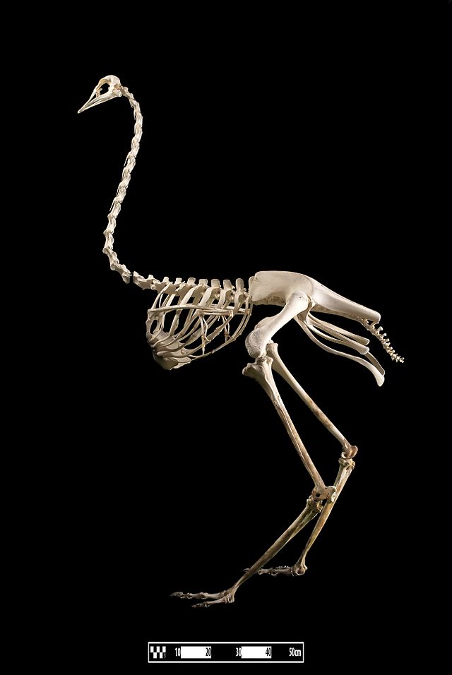
Figure 9.9 – Ostrich Skeleton © Museum of Veterinary Anatomy FMVZ USP is licensed under a CC BY (Attribution) license
Figure 9.10 – Limpkin Ribs © REM is licensed under a Public Domain license
Figure 9.11 – Human Ribs and Sternum © OpenStax is licensed under a CC BY (Attribution) license
Figure 9.12 – Amphioxus © OpenStax is licensed under a CC BY (Attribution) license
Figure 9.13 – Fish vertebrae © Bill Ryerson is licensed under a CC BY (Attribution) license
Figure 9.14 – Modified human atlas © OpenStax is licensed under a CC BY (Attribution) license
Figure 9.15 – Frog Skeleton © Lambert et al 2017: https://doi.org/10.3897/zse.93.10188 is licensed under a CC BY (Attribution) license
Figure 9.16 – Snake-necked turtle © Marijan Tunjic is licensed under a CC BY (Attribution) license
Figure 9.17 – Turtle skeleton © Kelsey Murdock is licensed under a CC BY (Attribution) license
Figure 9.18 – Bird skeletons © Christian Heinrich Pander and Eduard d’ Alton is licensed under a CC BY (Attribution) license
Figure 9.19 – Bird skeleton © Jrockley is licensed under a CC BY-SA (Attribution ShareAlike) license
Figure 9.20 – Human vertebral column © OpenStax is licensed under a CC BY (Attribution) license
Figure 9.21 – Giraffe and blue whale vertbrae © Bill Ryerson is licensed under a CC BY (Attribution) license
Figure 9.22 – Cheetah gallop © Dennis Donohue is licensed under a CC BY (Attribution) license
Figure 9.23 – Human herniated disc © SMART-Servier Medical Art, part of Laboratoires Servier. is licensed under a CC BY (Attribution) license
Chapter 10
Figure 10.1 – Skeleton differences © OpenStax is licensed under a CC BY-SA (Attribution ShareAlike) license
Figure 10.2 – Fin-fold vs. Gill-arch theory © Vanessa K Hilliard is licensed under a CC BY (Attribution) license
Figure 10.3 – Fin structure © Vanessa K Hilliard is licensed under a CC BY (Attribution) license
Figure 10.4 – ancestral relative position of the pelvis to the vertebral column © Vanessa K Hilliard is licensed under a CC BY (Attribution) license
Figure 10.5 – pectoral girdle structure © Vanessa K Hilliard is licensed under a CC BY (Attribution) license
Figure 10.6 – fin vs limb structure © Vanessa K Hilliard is licensed under a CC BY (Attribution) license
Figure 10.7 – fin vs limb developmental axis © Vanessa K Hilliard is licensed under a CC BY (Attribution) license
Figure 10.8 – three early tetrapods is licensed under a CC BY (Attribution) license
Figure 10.9 – early tetrapod pectoral and pelvic girdles © Vanessa K Hilliard is licensed under a CC BY (Attribution) license
Figure 10.10 – girdle attachment © Vanessa K Hilliard is licensed under a CC BY (Attribution) license
Figure 10.11 – dog pelvis is licensed under a Public Domain license
Figure 10.12 – human pelvis © U.S. National Cancer Institute, Fred the Oyster is licensed under a CC BY-SA (Attribution ShareAlike) license
Figure 10.13 – armadillo arm © David Stang is licensed under a CC BY-SA (Attribution ShareAlike) license
Figure 10.14 – astragalus © BodyParts3D is made by DBCLS is licensed under a CC BY-SA (Attribution ShareAlike) license
Figure 10.15 – ichthyosaur paddle © Samuel Wendell Williston is licensed under a Public Domain license
Figure 10.16 – wings © John Romanes is licensed under a Public Domain license
Figure 10.17 – bird skeleton © Vanessa K Hilliard is licensed under a CC BY (Attribution) license
Figure 10.18 – synapsid vs mammal pectoral girdle © Vanessa K Hilliard is licensed under a CC BY (Attribution) license
Figure 10.19 – human scapula © Wikimedia is licensed under a CC BY-SA (Attribution ShareAlike) license
Figure 10.20 – human femur © Anatomography is licensed under a CC BY-SA (Attribution ShareAlike) license
Figure 10.21 – human forearm © OpenStax is licensed under a CC BY-SA (Attribution ShareAlike) license
Figure 10.22 – human knee © Patrick J. Lynch, medical illustrator is licensed under a CC BY (Attribution) license
Chapter 11
Figure 11.1 – muscle tissue types © Vanessa K Hilliard is licensed under a CC BY (Attribution) license
Figure 11.2 – myocyte © Vanessa K Hilliard is licensed under a CC BY (Attribution) license
Figure 11.3 – sarcomere and filaments © Betts et al. is licensed under a CC BY (Attribution) license
Figure 11.4 – cusk eel © Betts et al. is licensed under a CC BY (Attribution) license
Figure 11.5 – sarcomere contraction © Betts et al. is licensed under a CC BY (Attribution) license
Figure 11.6 – cusk eel © NOAA via @DeepSeaImage. is licensed under a Public Domain license
Figure 11.7 – NMJ © Betts et al. is licensed under a CC BY (Attribution) license
Figure 11.8 – antagonist muscles © Vanessa K Hilliard is licensed under a CC BY (Attribution) license
Figure 11.9 – fused vs unfused tetanus © Vanessa K Hilliard is licensed under a CC BY (Attribution) license
Figure 11.10 – length tension relationship © Daniel Walsh and Alan Sved; Vanessa K Hilliard is licensed under a CC BY (Attribution) license
Figure 11.11 – muscle fiber types © Betts et al. is licensed under a CC BY (Attribution) license
Chapter 12
Figure 12.1 – muscle organization © Betts et al. is licensed under a CC BY-SA (Attribution ShareAlike) license
Figure 12.2 – tendon anatomy © Vanessa K Hilliard is licensed under a CC BY (Attribution) license
Figure 12.3 – origin vs insertion © Vanessa K Hilliard is licensed under a CC BY (Attribution) license
Figure 12.4 – muscle shapes © OpenStax College is licensed under a CC BY-SA (Attribution ShareAlike) license
Figure 12.5 – shark vs. human extraocculars © Vanessa K Hilliard is licensed under a CC BY (Attribution) license
Figure 12.6 – jaw closing muscles © Vanessa K Hilliard is licensed under a CC BY (Attribution) license
Figure 12.7 – body wall layers © Vanessa K Hilliard is licensed under a CC BY (Attribution) license
Figure 12.8 – human musculature © OpenStax College is licensed under a CC BY (Attribution) license
Figure 12.9 – human muscular anatomy © Betts et al. is licensed under a CC BY-SA (Attribution ShareAlike) license
Figure 12.10 – piriformis © John Kiel, Chris Hauglid
Figure 12.11 – iliopsoas © OpenStax College is licensed under a CC BY (Attribution) license
Chapter 13
Figure 13.1 – Vertebreate gut © Donovan P. German is licensed under a CC BY (Attribution) license
Figure 13.2 – Mammalian stomach © Stevens CE, Hume ID
Figure 13.3 – Intestine © NIH / National Cancer Institute is licensed under a Public Domain license
Figure 13.4 – Anatomy of Intestine © Donovan P. German is licensed under a CC BY (Attribution) license
Figure 13.5 – Digestive system of fish © OpenStax College is licensed under a CC BY (Attribution) license
Figure 13.6 – Principle nutrients © Donovan P. German is licensed under a CC BY (Attribution) license
Figure 13.7 – Nominal surface area of small intestines © Caviedes-Vidal, E, et. al (Non-commercial use permitted per https://doi.org/10.1073/pnas.0405514101)
Figure 13.8 – Synteny maps © Donovan P. German is licensed under a CC BY (Attribution) license
Chapter 14
Figure 14.1 – Diffusion animation © Jacopo Bertolotti is licensed under a CC0 (Creative Commons Zero) license
Figure 14.2 – Scaling relationship © Elska Kaczmarek, Jackson Phillips, and Bill Ryerson is licensed under a CC BY (Attribution) license
Figure 14.3 – Basic respiratory anatomy © Elska Kaczmarek, Jackson Phillips, and Bill Ryerson is licensed under a CC BY (Attribution) license
Figure 14.4 – Ventilation pump mechanisms © Samuel Garman is licensed under a CC0 (Creative Commons Zero) license
Figure 14.5 – frog skin folds © Elska Kaczmarek, Jackson Phillips, and Bill Ryerson is licensed under a CC BY (Attribution) license
Figure 14.6 – Lung budding development © OpenStax is licensed under a CC BY-SA (Attribution ShareAlike) license
Figure 14.7 – Salamander egg mass © Alpinetrout is licensed under a CC BY-SA (Attribution ShareAlike) license
Figure 14.8 – Embryos © Ross, C. and Boroviak, T.E is licensed under a CC BY (Attribution) license
Figure 14.9 – Respiration phylogeny © Levi; Phạm Thanh Lộc; sachin modgekar; Elska Kaczmarek, Jackson Phillips, and Bill Ryerson is licensed under a CC BY (Attribution) license
Figure 14.10 – Respiration diversity and ecology © Elska Kaczmarek, Jackson Phillips, and Bill Ryerson is licensed under a CC BY (Attribution) license
Figure 14.11 – Gill anatomy © Elska Kaczmarek, Jackson Phillips, and Bill Ryerson is licensed under a CC BY (Attribution) license
Figure 14.12 – lung anatomy © Elska Kaczmarek, Jackson Phillips, and Bill Ryerson is licensed under a CC BY (Attribution) license
Figure 14.13 – 2- and 4-stroke breaths © Elska Kaczmarek, Jackson Phillips, and Bill Ryerson is licensed under a CC BY (Attribution) license
Figure 14.14 – Lung chambers © Elska Kaczmarek, Jackson Phillips, and Bill Ryerson is licensed under a CC BY (Attribution) license
Figure 14.15 – crocodile diaphragmaticus © Elska Kaczmarek, Jackson Phillips, and Bill Ryerson is licensed under a CC BY (Attribution) license
Figure 14.16 – bird ventilation © Elska Kaczmarek, Jackson Phillips, and Bill Ryerson is licensed under a CC BY (Attribution) license
Figure 14.17 – Human lungs © Uwe Gille is licensed under a CC BY-SA (Attribution ShareAlike) license
Figure 14.18 – mammal ventilation © OpenStax is licensed under a CC BY-SA (Attribution ShareAlike) license
Figure 14.19 – human lung branching © OpenStax is licensed under a CC BY-SA (Attribution ShareAlike) license
Figure 14.20 – intrapleural space © OpenStax is licensed under a CC BY-SA (Attribution ShareAlike) license
Figure 14.21 – human lung capacities © Laboratoires Servier is licensed under a CC BY-SA (Attribution ShareAlike) license
Figure 14.22 – Branching pattern © OpenStax is licensed under a CC BY-SA (Attribution ShareAlike) license
Figure 14.23 – Human lungs and pleural cavity © OpenStax is licensed under a CC BY-SA (Attribution ShareAlike) license
Figure 14.24 © Johnson, A.E.W., Pollard, T.J., Berkowitz, S.J. et al. is licensed under a CC BY-SA (Attribution ShareAlike) license
Figure 14.25 – Chest X-Ray © OpenStax is licensed under a CC BY-SA (Attribution ShareAlike) license
Figure 14.26 – Lung volumes and capacities © OpenStax is licensed under a CC BY-SA (Attribution ShareAlike) license
Chapter 15
Figure 15.1 – Circulation schematic © Bjarke Jensen, William Joyce, Tobias Wang, Bill Ryerson, and Lisa Whitenack is licensed under a CC BY (Attribution) license
Figure 15.2 – Artery Structure © Blausen.com staff is licensed under a CC BY (Attribution) license
Figure 15.3 – Capillary type © Elizabeth2424 is licensed under a CC BY (Attribution) license
Figure 15.4 – Vessel blood pressure © Bjarke Jensen, William Joyce, Tobias Wang, Bill Ryerson, and Lisa Whitenack is licensed under a CC BY (Attribution) license
Figure 15.5 – Lymphatic capillaries © CFCF is licensed under a CC BY-SA (Attribution ShareAlike) license
Figure 15.6 – Lymphatic organs © Blausen.com staff is licensed under a CC BY (Attribution) license
Figure 15.7 – Embryo heart © Jenson et al. (Reused with permission per https://www.elsevier.com/about/policies-and-standards/copyright)
Figure 15.8 – Fish circulation © OpenStax is licensed under a CC BY (Attribution) license
Figure 15.9 – Pulmonary and systemic circuits © OpenStax is licensed under a CC BY (Attribution) license
Figure 15.10 – Aortic arches © Tatco V is licensed under a CC BY (Attribution) license
Figure 15.10 – Aortic arches © Tatco V is licensed under a CC BY (Attribution) license
Figure 15.11 – Vert hearts © Bjarke Jensen, William Joyce, Tobias Wang, Bill Ryerson, and Lisa Whitenack is licensed under a CC BY (Attribution) license
Figure 15.12 – Vert aortic arches © Bjarke Jensen, William Joyce, Tobias Wang, Bill Ryerson, and Lisa Whitenack is licensed under a CC BY (Attribution) license
Figure 15.13 – Heart cross-sections © Bjarke Jensen, William Joyce, Tobias Wang, Bill Ryerson, and Lisa Whitenack is licensed under a CC BY (Attribution) license
Figure 15.14 – Fish breathing schematics © Bjarke Jensen, William Joyce, Tobias Wang, Bill Ryerson, and Lisa Whitenack is licensed under a CC BY (Attribution) license
Figure 15.15 – Lungfish heart © Bjarke Jensen, William Joyce, Tobias Wang, Bill Ryerson, and Lisa Whitenack is licensed under a CC BY (Attribution) license
Figure 15.16 – Shunting © Bjarke Jensen, William Joyce, Tobias Wang, Bill Ryerson, and Lisa Whitenack is licensed under a CC BY (Attribution) license
Figure 15.17 – Amphibian heart © Jon Houseman is licensed under a CC BY-SA (Attribution ShareAlike) license
Figure 15.18 – Generalized reptile heart © Bjarke Jensen, William Joyce, Tobias Wang, Bill Ryerson, and Lisa Whitenack is licensed under a CC BY (Attribution) license
Figure 15.19 – Schematics of bloodflow © Bjarke Jensen, William Joyce, Tobias Wang, Bill Ryerson, and Lisa Whitenack is licensed under a CC BY (Attribution) license
Figure 15.20 – Crocodile Heart © LittleJerry is licensed under a CC BY-SA (Attribution ShareAlike) license
Figure 15.21 – Cardiac Architecture © Bjarke Jensen, William Joyce, Tobias Wang, Bill Ryerson, and Lisa Whitenack is licensed under a CC BY (Attribution) license
Figure 15.22 – Cardian Conduction © Panara, Virginia, Zuzana Varaliová, Jörg Wilting, Katarzyna Koltowska, and Michael Jeltsch is licensed under a CC BY (Attribution) license
Figure 15.23 – Vertebrate cladogram © Banda, C.H., Shiraishi, M., Mitsui, K. et al. is licensed under a CC BY (Attribution) license
Figure 15.24 – The secondary vascular system (SVS) and lymphatic vascular system (LVS) in ray-finned fishes © OpenStax is licensed under a CC BY (Attribution) license
Chapter 16
Figure 16.1 – General nephron © Lisa B. Whitenack is licensed under a CC BY (Attribution) license
Figure 16.2 – Bovine kidney capillaries © Wagner Souza e Silva / Museum of Veterinary Anatomy FMVZ USP is licensed under a CC BY-SA (Attribution ShareAlike) license
Figure 16.3 – Nephron particualrs © OpenStax Biology 2e is licensed under a CC BY-SA (Attribution ShareAlike) license
Figure 16.4 – Nephrotome drawing © NEW YORK HENRY HOLT AND COMPANY 1908 is licensed under a Public Domain license
Figure 16.5 – Tripartite kidney © Ashley Sawle is licensed under a CC BY-SA (Attribution ShareAlike) license
Figure 16.6 – Hagfish kidney © Lisa B. Whitenack is licensed under a CC BY (Attribution) license
Figure 16.7 – Lamprey life cycle © Salas CA, Yopak KE, Warrington RE, Hart NS, Potter IC and Collin SP is licensed under a CC BY (Attribution) license
Figure 16.8 – Shark kidney © Lisa B. Whitenack is licensed under a CC BY (Attribution) license
Figure 16.9 – Salmon © Hans-Petter Fjeld is licensed under a CC BY-SA (Attribution ShareAlike) license
Figure 16.10 – Bull shark © Pterantula is licensed under a CC BY-SA (Attribution ShareAlike) license
Figure 16.11 – Head kidney © Lisa B. Whitenack is licensed under a CC BY (Attribution) license
Figure 16.12 – Fish osmoreg © Kare Kare modified by Biezl AND Raver, Duane; modified by Biezl is licensed under a CC BY (Attribution) license
Figure 16.13 – Nephrons © Lisa B. Whitenack is licensed under a CC BY (Attribution) license
Figure 16.14 – Frog anatomy © Agha Haseebullah Khan is licensed under a CC BY-SA (Attribution ShareAlike) license
Figure 16.15 – Mammal kidneys © L. Keogh; D. Kilroy; S. Bhattacharjee is licensed under a CC BY (Attribution) license
Figure 16.16 – Human kidney © Open Stax is licensed under a CC BY (Attribution) license
Figure 16.17 – Human nephron © Jennifer Lange is licensed under a CC BY (Attribution) license
Figure 16.18 – Dolphin kidney © Sur, Roger L.; Meegan, Jenny M.; Smith, Cynthia R.; Schmitt, Todd; L’Esperance, James; Hendrikson, Dean; Woo, Jason R. is licensed under a CC BY (Attribution) license
Figure 16.19 – Snake anatomy © Uwe Gille is licensed under a CC BY-SA (Attribution ShareAlike) license
Figure 16.20 – Avian kidney © Lisa B. Whitenack is licensed under a CC BY (Attribution) license
Chapter 17
Figure 17.1 – General gonads © Kanamori & Kobayashi. is licensed under a CC BY (Attribution) license
Figure 17.2 – Rabbit ovary section © J. Bredl, RVC is licensed under a CC BY-NC-ND (Attribution NonCommercial NoDerivatives) license
Figure 17.3 – Ovary with broad ligament © Henry Gray is licensed under a Public Domain license
Figure 17.4 – Human testis © Openstax Human Anatomy & Physiology is licensed under a CC BY (Attribution) license
Figure 17.5 – Sex differentiation © Original work, adapted from Gilbert SF. is licensed under a CC BY (Attribution) license
Figure 17.6 – Oogenesis © Openstax Human Anatomy & Physiology is licensed under a CC BY (Attribution) license
Figure 17.7 – Sperm production © Openstax Human Anatomy & Physiology is licensed under a CC BY (Attribution) license
Figure 17.8 – Sequenetial hermaphrodism © Brooke Fitzwater (Bfitzwater) is licensed under a CC BY-SA (Attribution ShareAlike) license
Figure 17.9 – Hagfish eggs © NOAA is licensed under a Public Domain license
Figure 17.10 – River lamprey © Kanamori & Kobayashi is licensed under a CC BY (Attribution) license
Figure 17.11 – Generalized vert urogenital © Kanamori & Kobayashi. is licensed under a CC BY (Attribution) license
Figure 17.12 – Porbeagle male © NOAA is licensed under a Public Domain license
Figure 17.13 – Blacktip reef claspers © lwolfartist is licensed under a CC BY (Attribution) license
Figure 17.14 – Fish testes © Kanamori & Kobayashi is licensed under a CC BY (Attribution) license
Figure 17.15 – Urogential papilla © Peter van der Sluijs is licensed under a CC BY-SA (Attribution ShareAlike) license
Figure 17.16 – Guppies © Amy E. Deacon, Hideyasu Shimadzu, Maria Dornelas, Indar W. Ramnarine & Anne E. Magurran is licensed under a CC BY (Attribution) license
Figure 17.17 – Frog gonads © Maribel Méndez-Tepepa, Cuauhtémoc Morales-Cruz, Edelmira García-Nieto & Arely Anaya-Hernández is licensed under a CC BY (Attribution) license
Figure 17.18 – Salamander mating © Roger Culos is licensed under a CC BY-SA (Attribution ShareAlike) license
Figure 17.19 – Amplexus © Fredlyfish4 is licensed under a CC BY-SA (Attribution ShareAlike) license
Figure 17.20 – Tailed frog © Mokele is licensed under a CC BY-SA (Attribution ShareAlike) license
Figure 17.21 – Turtle penis © Jollie, Malcolm is licensed under a Public Domain license
Figure 17.22 – CT scan hemipenes © Roberts JR, Iova B, Austin CC is licensed under a CC BY (Attribution) license
Figure 17.23 – Rattlesnake hemipenes © Tess Thornton is licensed under a CC BY-SA (Attribution ShareAlike) license
Figure 17.24 – Lizard hemipenes © Sharp Photography, sharpphotography is licensed under a CC BY-SA (Attribution ShareAlike) license
Figure 17.25 – Gecko mating © Basile Morin is licensed under a CC BY (Attribution) license
Figure 17.26 – Mallard penis © Glen Bowman from Newcastle, England is licensed under a CC BY (Attribution) license
Figure 17.27 – Dove mating © Satdeep Gill is licensed under a CC BY-SA (Attribution ShareAlike) license
Figure 17.28 – Descent of testes is licensed under a Public Domain license
Figure 17.29 – Platypus anatomy male © Esouc32 is licensed under a CC BY-SA (Attribution ShareAlike) license
Figure 17.30 – Racoon baculum © Max Charping is licensed under a CC BY (Attribution) license
Figure 17.31 – shark ovaries © NOAA is licensed under a Public Domain license
Figure 17.32 – Teleost ovary stuff © Lisa B. Whitenack is licensed under a CC BY (Attribution) license
Figure 17.33 – Generalized vert urogenital © Kanamori & Kobayashi. is licensed under a CC BY (Attribution) license
Figure 17.34 – Materpiscus fossil © Sularko Museum Victoria Derivative work MagentaGreen is licensed under a CC BY-SA (Attribution ShareAlike) license
Figure 17.35 – Materpiscus reconstruction © Sularko is licensed under a CC BY-SA (Attribution ShareAlike) license
Figure 17.36 – Frog ovaries © Maribel Méndez-Tepepa, Cuauhtémoc Morales-Cruz, Edelmira García-Nieto & Arely Anaya-Hernández is licensed under a CC BY (Attribution) license
Figure 17.37 – Salamander eggs © Kent Mason/USFWS is licensed under a Public Domain license
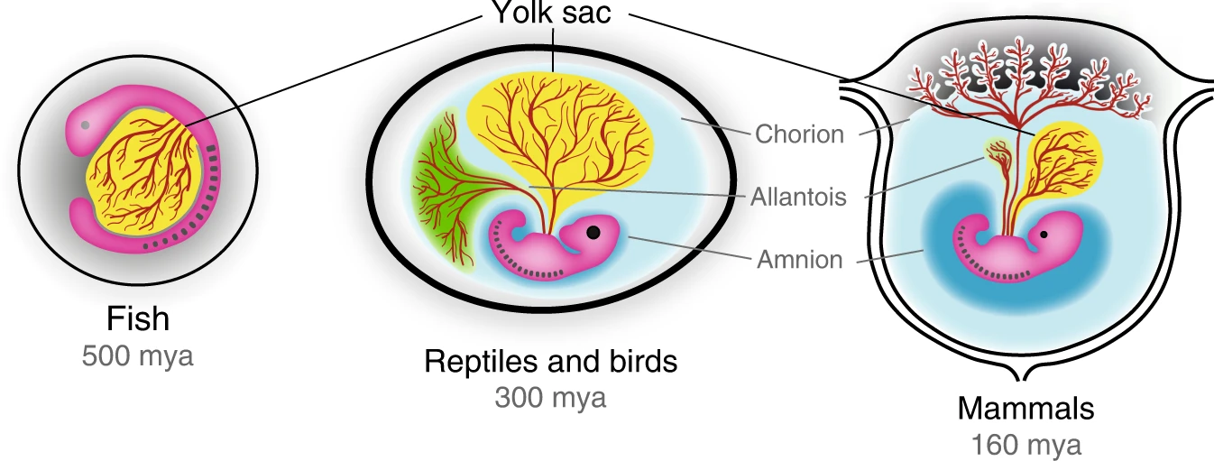
Figure 17.38 – Amniotic egg evolution © Ross, C., Boroviak, T.E. is licensed under a CC BY (Attribution) license
Figure 17.39 – Clitoris © Michael Schünke, Erik Schulte, Udo Schumacher is licensed under a CC0 (Creative Commons Zero) license
Figure 17.40 – Bird oviduct © Keyanapardilla98 is licensed under a CC BY-SA (Attribution ShareAlike) license
Figure 17.41 – bird vaginas © Patricia L.R. Brennan ,Richard O. Prum,Kevin G. McCracken,Michael D. Sorenson,Robert E. Wilson,Tim R. Birkhead is licensed under a CC BY (Attribution) license
Figure 17.42 – Snake ovary © Internet Archive Book Images is licensed under a Public Domain license
Figure 17.43 – Platypus anatomy male © Internet Archive Book Images is licensed under a Public Domain license
Figure 17.44 – Uteruses © Sciencia58 is licensed under a CC0 (Creative Commons Zero) license
Figure 17.45 – Mole bits © Rafael Jiménez1, Miguel Burgos1, and Francisco J. Barrionuevo1 is licensed under a CC BY (Attribution) license
Figure 17.46 – Spotted hyena © flowcomm is licensed under a CC BY (Attribution) license
Figure 17.47 – Spotted hyena reproductive tracts © Anatomische Gesellschaft is licensed under a Public Domain license
Figure 17.48 – Placenta © OpenStax College is licensed under a CC BY (Attribution) license
Figure 17.49 – Mammary glands: Mammal with nipples; Mammal with teets © Ruth Lawson is licensed under a CC BY (Attribution) license
Figure 17.50 – Human uterus © OpenStax College is licensed under a CC BY (Attribution) license
Figure 17.51 – Human vulva © OpenStax College is licensed under a CC BY (Attribution) license
Figure 17.52 – Human testes © OpenStax College is licensed under a CC BY (Attribution) license
Figure 17.53 – Human penis © OpenStax College is licensed under a CC BY (Attribution) license
Figure 17.54 – Birth canal © Stansfield, E., Fischer, B., Grunstra, N.D.S. et al. is licensed under a CC BY (Attribution) license
Figure 17.55 – Pelvis dimoprhism © Anatomy & Physiology is licensed under a CC BY (Attribution) license
Figure 17.56 – Q angle © Anatomy & Physiology is licensed under a CC BY-SA (Attribution ShareAlike) license
Chapter 18
Figure 18.1 – CNS vs PNS © OpenStax is licensed under a CC BY (Attribution) license
Figure 18.2 – Neuron © OpenStax is licensed under a CC BY (Attribution) license
Figure 18.3 – Brain lobes © OpenStax is licensed under a CC BY-SA (Attribution ShareAlike) license
Figure 18.4 – Glial cells © OpenStax is licensed under a CC BY (Attribution) license
Figure 18.5 – Brain development © OpenStax is licensed under a CC BY (Attribution) license
Figure 18.6 – Dogfish and human © Wikimedia user Looie496 is licensed under a CC BY (Attribution) license
Figure 18.7 – Ventricles © Wikimedia user BruceBlaus is licensed under a CC BY (Attribution) license
Figure 18.8 – Meninges © www.scientificanimations.com is licensed under a CC BY (Attribution) license
Figure 18.9 – Brain development © OpenStax is licensed under a CC BY (Attribution) license
Figure 18.10 – Spinal cords of four different tetrapod species © Bill Ryerson is licensed under a CC BY (Attribution) license
Figure 18.11 – Transverse sections of the spinal cord © Bill Ryerson is licensed under a CC BY (Attribution) license
Figure 18.12 – Variation of spinal cord structure in an ostrich © Bill Ryerson is licensed under a CC BY (Attribution) license
Figure 18.13 – Lumbar puncture © Cancer Research UK is licensed under a CC BY-SA (Attribution ShareAlike) license
Figure 18.14 – Eagle © White and Yellow Shallow Focus Bird Photography is licensed under a CC0 (Creative Commons Zero) license
Figure 18.15 – Lamprey Brain © Loonen, Anton JM, and Svetlana A. Ivanova. is licensed under a CC BY (Attribution) license
Figure 18.16 – Gray and white matter © OpenStax is licensed under a CC BY (Attribution) license
Figure 18.17 – Zebrafish brain © Bloch, Solal, Manon Thomas, Ingrid Colin, Sonya Galant, Elodie Machado, Pierre Affaticati, Arnim Jenett, and Kei Yamamoto. is licensed under a CC BY (Attribution) license
Figure 18.18 – Pallium © Wikimedia user Looie496 is licensed under a CC BY (Attribution) license
Figure 18.19 – Frog brain © Bill Ryerson is licensed under a CC BY (Attribution) license
Figure 18.20 – Alligator brain © Bill Ryerson is licensed under a CC BY (Attribution) license
Figure 18.21 – Wulst © Graham R. Martin , Kerry-Jayne Wilson, J. Martin Wild, Stuart Parsons, M. Fabiana Kubke, Jeremy Corfield is licensed under a CC BY (Attribution) license
Figure 18.22 – Mouse and human brain © Lauren N. Miterko, Elizabeth P. Lackey, Elizabeth P. Lackey, Detlef H. Heck, Roy V. Sillitoe is licensed under a CC BY (Attribution) license
Figure 18.23 – Mammal brains © University of Wisconsin and Michigan State Comparative Mammalian Brain Collections is licensed under a CC BY (Attribution) license
Figure 18.24 – Brain lobes © OpenStax is licensed under a CC BY (Attribution) license
Figure 18.25 – Cortices and association areas © OpenStax is licensed under a CC BY (Attribution) license
Figure 18.26 – Homunculus © OpenStax is licensed under a CC BY (Attribution) license
Chapter 19
Figure 19.1 – Sensory and motor pathways © Wikipedia user MartaAguayo is licensed under a CC BY-SA (Attribution ShareAlike) license
Figure 19.2 – Autonomic system in humans © Meredith Pomietlo is licensed under a CC BY-SA (Attribution ShareAlike) license
Figure 19.3 – Spinal nerve © Vectorized in CorelDraw by Mysid on an existing image at en-wiki by Tristanb. is licensed under a CC BY-SA (Attribution ShareAlike) license
Figure 19.4 – Cervical and brachial plexus © OpenStax.org is licensed under a CC BY-SA (Attribution ShareAlike) license
Figure 19.5 – Human cranial nerves © OpenStax.org is licensed under a CC BY-SA (Attribution ShareAlike) license
Figure 19.6 – Cranial nerve of sharks © Bill Ryerson is licensed under a CC BY (Attribution) license
Figure 19.7 – Laryngeal nerve © Bill Ryerson is licensed under a CC BY (Attribution) license
Chapter 20
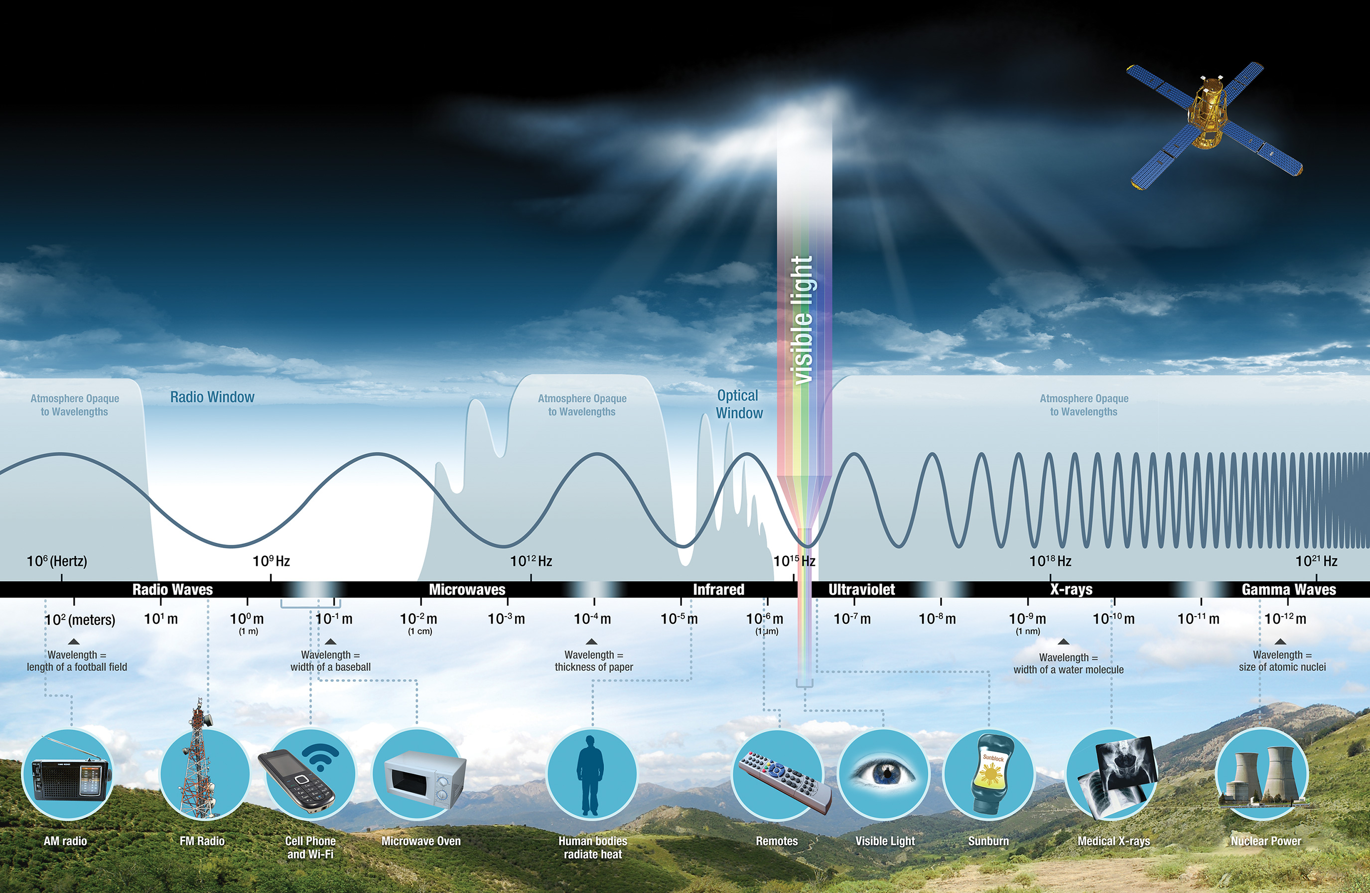
Figure 20.1 – Spectrum of electromagnetic radiation © NASA is licensed under a Public Domain license
Figure 20.2 – Primate eye and retina © OpenStax Human AP Book is licensed under a CC BY (Attribution) license
Figure 20.3 – Eye structure across vertebrates © Brett Aiello, Lauren Simonitis, Kate Criswell, and Bill Ryerson is licensed under a CC BY (Attribution) license
Figure 20.4 – Slit pupil © Brett Aiello, Lauren Simonitis, Kate Criswell, and Bill Ryerson is licensed under a CC BY (Attribution) license
Figure 20.5 – Development of vertebrate eyes © Brett Aiello, Lauren Simonitis, Kate Criswell, and Bill Ryerson is licensed under a CC BY (Attribution) license
Figure 20.6 – Main olfactory system © OpenStax Human AP Book is licensed under a CC BY (Attribution) license
Figure 20.7 – Human tongue papillae © OpenStax Human AP Book is licensed under a CC BY (Attribution) license
Figure 20.8 – Vestibular apparatus anatomy © OpenStax Human AP Book is licensed under a CC BY (Attribution) license
Figure 20.9 – Semicircular canals © OpenStax Human AP Book is licensed under a CC BY (Attribution) license
Figure 20.10 – Mammalian auditory system © OpenStax Human AP Book is licensed under a CC BY (Attribution) license
Figure 20.11 – Cross-sectional anatomy of the human cochlea © OpenStax Human AP Book is licensed under a CC BY (Attribution) license
Chapter 21
Figure 21.1 – Major endocrine glands © Jennifer L. Houtz is licensed under a CC BY (Attribution) license
Figure 21.2 – Exocrine and endocrine glands © Jennifer L. Houtz is licensed under a CC BY (Attribution) license
Figure 21.3 – Protein versus steroid hormones © BorisTM is licensed under a CC0 (Creative Commons Zero) license
Figure 21.4 – Negative feedback loop © Gordon Betts, Kelly A. Young, James A. Wise, Eddie Johnson, Brandon Poe, Dean H. Kruse, Oksana Korol, Jody E. Johnson, Mark Womble, Peter DeSaix is licensed under a CC BY (Attribution) license
Figure 21.5 – Hypothalamus and pituitary gland in a human © J. Gordon Betts, Kelly A. Young, James A. Wise, Eddie Johnson, Brandon Poe, Dean H. Kruse, Oksana Korol, Jody E. Johnson, Mark Womble, Peter DeSaix is licensed under a CC BY (Attribution) license
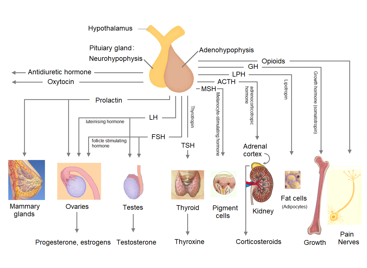
Figure 21.6 – Tropic hormones from the pituitary gland © Richard E. Jones, Kristin Lopez., is licensed under a CC BY-SA (Attribution ShareAlike) license
Figure 21.7 – Two lobes of pituitary gland © Jennifer L. Houtz is licensed under a CC BY (Attribution) license
Figure 21.8 – Pigeon crop milk © Ruggero Turra is licensed under a CC BY-SA (Attribution ShareAlike) license
Figure 21.9 – Hypothalamus evolution © Santiago-Andres, Golan and Fiordelisi is licensed under a CC BY (Attribution) license
Figure 21.10 – Pituitary arrangment © Jennifer L. Houtz is licensed under a CC BY (Attribution) license
Figure 21.11 – Human pineal gland © Vasey, McBride, and Penta is licensed under a CC BY (Attribution) license
Figure 21.12 – Parietal eye © TheAlphaWolf is licensed under a CC BY-SA (Attribution ShareAlike) license
Figure 21.13 – Hypothalamic-pituitary-thyroid (HPT) axis © Jennifer L. Houtz is licensed under a CC BY (Attribution) license
Figure 21.14 – Human thyroid and parathyroid glands © Jennifer L. Houtz is licensed under a CC BY (Attribution) license
Figure 21.15 – Regulation of blood calcium levels © Mkaram19 is licensed under a CC BY (Attribution) license
Figure 21.16 – Pancreatic islets © Jennifer L. Houtz is licensed under a CC BY (Attribution) license
Figure 21.17 – Blood glucose regulation © Carogonz11 is licensed under a CC BY-SA (Attribution ShareAlike) license
Figure 21.18 – Brockmann body (Figure 2) from Chanet et al. (2023), Scientific Reports © Chanet, Schnell, Guintard, Chen is licensed under a CC BY (Attribution) license
Figure 21.19 – Adrenal gland sitting on top of kidney showing medulla vs. cortex layers © Jennifer L. Houtz is licensed under a CC BY (Attribution) license
Figure 21.20 – Hypothalamic-pituitary-adrenal (HPA axis) © Jennifer L. Houtz is licensed under a CC BY (Attribution) license
Figure 21.21 – Adrenal glands across Squamata (Figure 1) from Capaldo 2023 Animals © Capaldo is licensed under a CC BY (Attribution) license
Figure 21.22 – Testes and ovaries © Jennifer L. Houtz is licensed under a CC BY (Attribution) license
Figure 21.23 – Examples of secondary sex characteristics © Adrian Pingstone; Karl Moor; Muñoz RC, Zgliczynski BJ, Laughlin JL, Teer BZ is licensed under a CC BY (Attribution) license
Figure 21.24 – Hypothalamic–pituitary-gonadal axis (HPG axis) © Jennifer L. Houtz is licensed under a CC BY (Attribution) license

_LACMA_M.87.271a-g_(6_of_8).jpg)
.jpg%20AND%20https://commons.wikimedia.org/wiki/File:Salp.jpg)
_(15149695488).jpg)
.jpg)
_1.jpg)
.svg)
.jpg)
_foot.jpg)
_2.jpg)
.jpg)
.jpg)
_(1933).jpg)
.JPG)
_male_and_female.png)
_(20585824866).jpg)
_Figure_3.jpg)
_male,_with_everted_hemipenes.jpg)
%22_(1866)_(14755631195).jpg)
_(20633924018).jpg)
_(18006271698).jpg)